Monday Poster Session
Category: Colon
P1981 - Unique Presentation of Colonic Single Stripe Sign
Monday, October 28, 2024
10:30 AM - 4:00 PM ET
Location: Exhibit Hall E

Has Audio

Sanya Badar, MD
The Wright Center for Graduate Medical Education
Scranton, PA
Presenting Author(s)
Sanya Badar, MD1, Saba Altarawneh, MD1, Mazen Tolaymat, MD2
1The Wright Center for Graduate Medical Education, Scranton, PA; 2Wilkes-Barre General Hospital, Scranton, PA
Introduction: Ischemic colitis (IC) characterized by hypoperfusion of the colon, can present uniquely with a colonic single stripe sign, which is pathognomonic for the condition. Ischemic colitis can manifest itself in a mild temporary form, which is why the incidence of the disease is significantly underestimated. This report highlights an unusual presentation of ischemic colitis in an elderly male patient.
Case Description/Methods: An 87-year-old male with a complex medical history, treated with radiation for prostate cancer presented with lower abdominal pain and non-bloody diarrhea for 2 days. Notably, he denied experiencing nausea, vomiting, hematochezia, or melena, and there was no history of recent antibiotic use or exposure to sickness. The patient was hemodynamically stable, routine laboratory reports were normal except for leukocytosis. Abdominal computed tomography confirmed the presence of colonic diverticula with mural thickening involving the descending and sigmoid colon, loss of haustral with increased peri colonic mesenteric stranding with tiny amount of fluid in the field suggestive of colitis. Colonoscopy showed severe mucosal friability 30-70 cm from the anal margin and identified the long linear ulceration known as “colonic single stripe” sign suggestive of IC. Endoscopic biopsies revealed cryptitis, enzootic necrotizing colitis, and focal fibrosis in the lamina propria of the colonic mucosa. In light of the clinical history, endoscopic, and histologic findings the patient was presumed to have IC, and discharged on oral antibiotics with GI follow-up. Two months later he was noted to be asymptomatic and planned repeat colonoscopy to ensure mucosal healing.
Discussion: The single stripe sign is a critical diagnostic marker favored mild IC. This sign can be attributed to the segmental nature of the involvement of the colon which would be consistent with IC since the lesion resembles linear ulceration that runs longitudinally along the colon and the length of the colon is usually related to the distribution of blood supply. This segmental vascular compromise at the mucosal level occurs due to ischemia or small vascular inadequacy. It has been inferred that linear demarcation explains the involvement of the mesenteric vascular border. This sign's finding underlines the value of appreciating typical imaging characteristics that define specific conditions to facilitate subsequent workup and management plans. Prompt recognition and management can lead to favorable outcomes in patients with IC.

Disclosures:
Sanya Badar, MD1, Saba Altarawneh, MD1, Mazen Tolaymat, MD2. P1981 - Unique Presentation of Colonic Single Stripe Sign, ACG 2024 Annual Scientific Meeting Abstracts. Philadelphia, PA: American College of Gastroenterology.
1The Wright Center for Graduate Medical Education, Scranton, PA; 2Wilkes-Barre General Hospital, Scranton, PA
Introduction: Ischemic colitis (IC) characterized by hypoperfusion of the colon, can present uniquely with a colonic single stripe sign, which is pathognomonic for the condition. Ischemic colitis can manifest itself in a mild temporary form, which is why the incidence of the disease is significantly underestimated. This report highlights an unusual presentation of ischemic colitis in an elderly male patient.
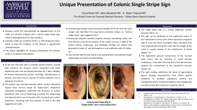
Case Description/Methods: An 87-year-old male with a complex medical history, treated with radiation for prostate cancer presented with lower abdominal pain and non-bloody diarrhea for 2 days. Notably, he denied experiencing nausea, vomiting, hematochezia, or melena, and there was no history of recent antibiotic use or exposure to sickness. The patient was hemodynamically stable, routine laboratory reports were normal except for leukocytosis. Abdominal computed tomography confirmed the presence of colonic diverticula with mural thickening involving the descending and sigmoid colon, loss of haustral with increased peri colonic mesenteric stranding with tiny amount of fluid in the field suggestive of colitis. Colonoscopy showed severe mucosal friability 30-70 cm from the anal margin and identified the long linear ulceration known as “colonic single stripe” sign suggestive of IC. Endoscopic biopsies revealed cryptitis, enzootic necrotizing colitis, and focal fibrosis in the lamina propria of the colonic mucosa. In light of the clinical history, endoscopic, and histologic findings the patient was presumed to have IC, and discharged on oral antibiotics with GI follow-up. Two months later he was noted to be asymptomatic and planned repeat colonoscopy to ensure mucosal healing.
Discussion: The single stripe sign is a critical diagnostic marker favored mild IC. This sign can be attributed to the segmental nature of the involvement of the colon which would be consistent with IC since the lesion resembles linear ulceration that runs longitudinally along the colon and the length of the colon is usually related to the distribution of blood supply. This segmental vascular compromise at the mucosal level occurs due to ischemia or small vascular inadequacy. It has been inferred that linear demarcation explains the involvement of the mesenteric vascular border. This sign's finding underlines the value of appreciating typical imaging characteristics that define specific conditions to facilitate subsequent workup and management plans. Prompt recognition and management can lead to favorable outcomes in patients with IC.

Figure: Colonoscopy imaging demonstrating "Single-stripe" sign in Descending Colon
Disclosures:
Sanya Badar indicated no relevant financial relationships.
Saba Altarawneh indicated no relevant financial relationships.
Mazen Tolaymat indicated no relevant financial relationships.
Sanya Badar, MD1, Saba Altarawneh, MD1, Mazen Tolaymat, MD2. P1981 - Unique Presentation of Colonic Single Stripe Sign, ACG 2024 Annual Scientific Meeting Abstracts. Philadelphia, PA: American College of Gastroenterology.
