Monday Poster Session
Category: Colon
P1982 - An Atypical Colon Polyp Leading to Diagnosis of Metastatic Breast Cancer
Monday, October 28, 2024
10:30 AM - 4:00 PM ET
Location: Exhibit Hall E

Has Audio
- SP
Sandesh R. Parajuli, MD
Reading Hospital - Tower Health
Reading, PA
Presenting Author(s)
Sandesh Parajuli, MD1, Daniel J. Rowan, MD2, Amna Iftikhar, MBBS3, Humberto Sifuentes, MD4, Asad Jehangir, MD4
1Reading Hospital - Tower Health, Reading, PA; 2North Shore Pathologists, Milwaukee, WI; 3Rawalpindi Medical University, Rawalpindi, Punjab, Pakistan; 4Augusta University, Augusta, GA
Introduction: Breast cancer is the most prevalent cancer among women worldwide. Every year it
accounts for one in three cancer diagnoses in females in the United States. The
common sites for breast cancer metastasis include brain, bones, lungs, and liver, but
metastasis to the gastrointestinal (GI) tract is uncommon. Here, we report a case of
metastatic breast cancer diagnosed after an atypical "polyp" was removed during
colonoscopy.
Case Description/Methods: An 87-year-old female with coronary artery disease, peripheral vascular disease on dual
anti platelet therapy, and adenomatous polyp on colonoscopy 5 years prior, presented with
fatigue. She appeared pale and was found to have normocytic anemic with hemoglobin
of 7.9 g/dl (12-14 g/dl), and mean corpuscular volume of 92.2 fL (80-100 fL). Her folate
level was low and iron studies showed borderline low iron level of 33, iron saturation of
33% and ferritin of 33.5. Esophagogastroduodenoscopy did not show any findings to
explain the anemia. Colonoscopy revealed a 12 mm colonic atypical polypoid lesion in
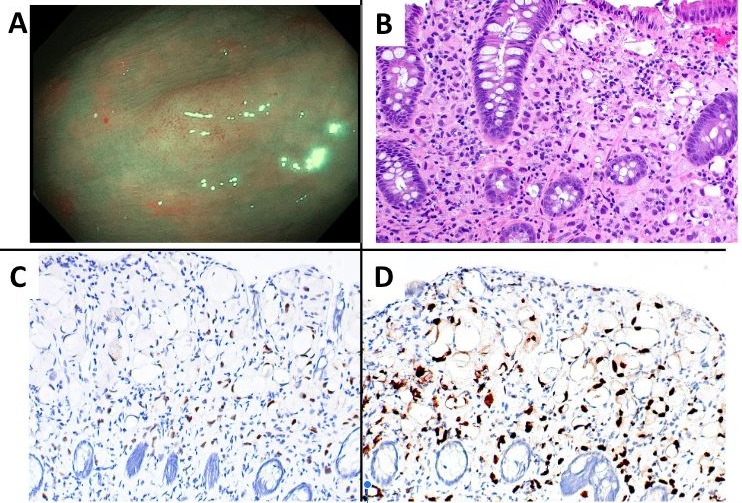
the ascending colon that was resected via cold snare.Pathology of the colon polyp showed adenocarcinoma involving the lamina propria. It was positive for keratin, AE1/AE3, CK7, GATA3, GCDFP-15, and Mammaglobin (focal).
These findings were suggestive of adenocarcinoma of breast which was ER +ve, PR -
ve and HER2 -ve on immunohistology. Patient’s last mammogram was 7 years prior
which showed a 5 mm irregular soft tissue density on her right breast, but she had
opted to not have it biopsied at that time. Her CA 27.29 level was elevated to 92.2 U/ml.
PET scan did not show any hypermetabolic activity. She then had ultrasound and
biopsy of the breast mass which was consistent with breast adenocarcinoma breast with
immunoprofile similar to that of the colonic polyp (ER +, PR -ve and HER2 -ve). Bone
marrow biopsy showed metastasis.Different treatment options
were discussed by oncology, and the patient decided to proceed with ribociclib.
Discussion: Colon polypectomy specimens leading to diagnosis of metastatic colon cancer is rare. Colon
polyps are sometimes not resected in elderly patients during inpatient colonoscopies
performed to investigate the cause of anemia, for fear of causing GI bleeding and
confound the clinical picture. Gastroenterologists should have a low threshold to sample
atypical appearing colon lesions, as this could lead to unexpected diagnosis.

Disclosures:
Sandesh Parajuli, MD1, Daniel J. Rowan, MD2, Amna Iftikhar, MBBS3, Humberto Sifuentes, MD4, Asad Jehangir, MD4. P1982 - An Atypical Colon Polyp Leading to Diagnosis of Metastatic Breast Cancer, ACG 2024 Annual Scientific Meeting Abstracts. Philadelphia, PA: American College of Gastroenterology.
1Reading Hospital - Tower Health, Reading, PA; 2North Shore Pathologists, Milwaukee, WI; 3Rawalpindi Medical University, Rawalpindi, Punjab, Pakistan; 4Augusta University, Augusta, GA
Introduction: Breast cancer is the most prevalent cancer among women worldwide. Every year it
accounts for one in three cancer diagnoses in females in the United States. The
common sites for breast cancer metastasis include brain, bones, lungs, and liver, but
metastasis to the gastrointestinal (GI) tract is uncommon. Here, we report a case of
metastatic breast cancer diagnosed after an atypical "polyp" was removed during
colonoscopy.
Case Description/Methods: An 87-year-old female with coronary artery disease, peripheral vascular disease on dual
anti platelet therapy, and adenomatous polyp on colonoscopy 5 years prior, presented with
fatigue. She appeared pale and was found to have normocytic anemic with hemoglobin
of 7.9 g/dl (12-14 g/dl), and mean corpuscular volume of 92.2 fL (80-100 fL). Her folate
level was low and iron studies showed borderline low iron level of 33, iron saturation of
33% and ferritin of 33.5. Esophagogastroduodenoscopy did not show any findings to
explain the anemia. Colonoscopy revealed a 12 mm colonic atypical polypoid lesion in
the ascending colon that was resected via cold snare.Pathology of the colon polyp showed adenocarcinoma involving the lamina propria. It was positive for keratin, AE1/AE3, CK7, GATA3, GCDFP-15, and Mammaglobin (focal).
These findings were suggestive of adenocarcinoma of breast which was ER +ve, PR -
ve and HER2 -ve on immunohistology. Patient’s last mammogram was 7 years prior
which showed a 5 mm irregular soft tissue density on her right breast, but she had
opted to not have it biopsied at that time. Her CA 27.29 level was elevated to 92.2 U/ml.
PET scan did not show any hypermetabolic activity. She then had ultrasound and
biopsy of the breast mass which was consistent with breast adenocarcinoma breast with
immunoprofile similar to that of the colonic polyp (ER +, PR -ve and HER2 -ve). Bone
marrow biopsy showed metastasis.Different treatment options
were discussed by oncology, and the patient decided to proceed with ribociclib.
Discussion: Colon polypectomy specimens leading to diagnosis of metastatic colon cancer is rare. Colon
polyps are sometimes not resected in elderly patients during inpatient colonoscopies
performed to investigate the cause of anemia, for fear of causing GI bleeding and
confound the clinical picture. Gastroenterologists should have a low threshold to sample
atypical appearing colon lesions, as this could lead to unexpected diagnosis.

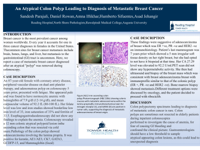
Figure: Figure (A)12 mm ascending colon polyp.Photomicrograph (H&E 200x) showing colonic mucosa with metastatic adenocarcinoma within the lamina propria(B), immunohistochemical stain for estrogen receptor (C), and GATA3 (D) are positive in tumor cells, supporting the diagnosis of metastatic adenocarcinoma of breast origin.
Disclosures:
Sandesh Parajuli indicated no relevant financial relationships.
Daniel Rowan indicated no relevant financial relationships.
Amna Iftikhar indicated no relevant financial relationships.
Humberto Sifuentes indicated no relevant financial relationships.
Asad Jehangir indicated no relevant financial relationships.
Sandesh Parajuli, MD1, Daniel J. Rowan, MD2, Amna Iftikhar, MBBS3, Humberto Sifuentes, MD4, Asad Jehangir, MD4. P1982 - An Atypical Colon Polyp Leading to Diagnosis of Metastatic Breast Cancer, ACG 2024 Annual Scientific Meeting Abstracts. Philadelphia, PA: American College of Gastroenterology.
