Monday Poster Session
Category: Colon
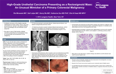
P2025 - High-Grade Urothelial Carcinoma Presenting as a Rectosigmoid Mass: An Unusual Mimicker of a Primary Colorectal Malignancy
Monday, October 28, 2024
10:30 AM - 4:00 PM ET
Location: Exhibit Hall E

Has Audio

Ria Minawala, MD
NYU Langone Health
New York, NY
Presenting Author(s)
Ria Minawala, MD, Saif Laljee, MD, Henry Wu, MD, Katherine Sun, MD, Elie Al Kazzi, MD, MPH
NYU Langone Health, New York, NY
Introduction: Urothelial carcinoma of the bladder typically metastasizes to lymph nodes, bones, lung, liver, and peritoneum; spread to the gastrointestinal tract is exceedingly rare. We report a case of a high-grade urothelial carcinoma presenting as a rectosigmoid mass, an exceptionally rare presentation of this malignancy.
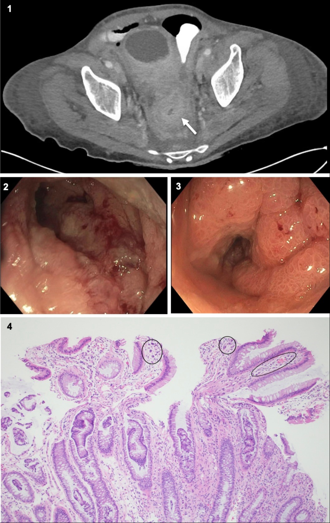
Case Description/Methods: A 67-year-old male with a history of Parkinson’s disease, depression, and chronic constipation presented from a subacute rehabilitation facility with a two-month history of decreased oral intake, nausea, abdominal pain, weakness, and unintentional weight loss. He denied obstructive symptoms and review of systems was normal. Of note, during a recent hospital admission for similar symptoms, CT abdomen and pelvis demonstrated circumferential thickening of the bladder and rectosigmoid, which was thought to represent stercoral colitis in the setting of chronic constipation. He was hemodynamically stable and physical exam was notable for diffuse abdominal tenderness and distension as well as bilateral lower extremity swelling. Labs were remarkable for normocytic anemia, hypoalbuminemia, and elevated carcinoembryonic antigen to 6.3 ng/ml. CT chest demonstrated a pericardial effusion, bilateral pleural effusions, and bilateral subsegmental pulmonary emboli. CT abdomen and pelvis revealed moderate bilateral hydronephrosis, marked thickening of the bladder and rectosigmoid (Figure 1) concerning for an underlying malignancy, and bilateral femoral deep venous thrombosis. A colonoscopy was performed, revealing a semi-circumferential and partially obstructing mass extending from the distal rectum to the rectosigmoid (Figure 2, 3). The scope was unable to traverse past the sigmoid colon due to tethering from the lesion. Biopsies were obtained of the approximately twelve-centimeter mass. Histopathological examination confirmed poorly differentiated carcinoma with neoplastic cells strongly positive for CK7, GATA 2, and uroplakin II, confirming a primary urinary bladder cancer (Figure 4). After goals of care discussion, systemic treatment was deferred, and decision was made to pursue hospice care.
Discussion: This was a case of metastatic urothelial carcinoma discovered in the rectosigmoid, an unusual mimicker of a primary colorectal malignancy. It is postulated that rectal involvement occurs by direct invasion of cancer cells. In patients who develop rectal constriction secondary to metastatic disease, a prognosis of less than one year has been reported.

Disclosures:
Ria Minawala, MD, Saif Laljee, MD, Henry Wu, MD, Katherine Sun, MD, Elie Al Kazzi, MD, MPH. P2025 - High-Grade Urothelial Carcinoma Presenting as a Rectosigmoid Mass: An Unusual Mimicker of a Primary Colorectal Malignancy, ACG 2024 Annual Scientific Meeting Abstracts. Philadelphia, PA: American College of Gastroenterology.
NYU Langone Health, New York, NY
Introduction: Urothelial carcinoma of the bladder typically metastasizes to lymph nodes, bones, lung, liver, and peritoneum; spread to the gastrointestinal tract is exceedingly rare. We report a case of a high-grade urothelial carcinoma presenting as a rectosigmoid mass, an exceptionally rare presentation of this malignancy.
Case Description/Methods: A 67-year-old male with a history of Parkinson’s disease, depression, and chronic constipation presented from a subacute rehabilitation facility with a two-month history of decreased oral intake, nausea, abdominal pain, weakness, and unintentional weight loss. He denied obstructive symptoms and review of systems was normal. Of note, during a recent hospital admission for similar symptoms, CT abdomen and pelvis demonstrated circumferential thickening of the bladder and rectosigmoid, which was thought to represent stercoral colitis in the setting of chronic constipation. He was hemodynamically stable and physical exam was notable for diffuse abdominal tenderness and distension as well as bilateral lower extremity swelling. Labs were remarkable for normocytic anemia, hypoalbuminemia, and elevated carcinoembryonic antigen to 6.3 ng/ml. CT chest demonstrated a pericardial effusion, bilateral pleural effusions, and bilateral subsegmental pulmonary emboli. CT abdomen and pelvis revealed moderate bilateral hydronephrosis, marked thickening of the bladder and rectosigmoid (Figure 1) concerning for an underlying malignancy, and bilateral femoral deep venous thrombosis. A colonoscopy was performed, revealing a semi-circumferential and partially obstructing mass extending from the distal rectum to the rectosigmoid (Figure 2, 3). The scope was unable to traverse past the sigmoid colon due to tethering from the lesion. Biopsies were obtained of the approximately twelve-centimeter mass. Histopathological examination confirmed poorly differentiated carcinoma with neoplastic cells strongly positive for CK7, GATA 2, and uroplakin II, confirming a primary urinary bladder cancer (Figure 4). After goals of care discussion, systemic treatment was deferred, and decision was made to pursue hospice care.
Discussion: This was a case of metastatic urothelial carcinoma discovered in the rectosigmoid, an unusual mimicker of a primary colorectal malignancy. It is postulated that rectal involvement occurs by direct invasion of cancer cells. In patients who develop rectal constriction secondary to metastatic disease, a prognosis of less than one year has been reported.

Figure: Figure 1: CTAP demonstrating marked thickening of the rectosigmoid.
Figure 2: Endoscopic view of mass extending into the sigmoid colon.
Figure 3: Endoscopic view of semi-circumferential rectal mass.
Figure 4: Rectal biopsy histopathology demonstrates poorly differentiated carcinoma along with discohesive cells with plasmacytoid features infiltrating the lamina propria, muscularis mucosa, and submucosa.
Disclosures:
Ria Minawala indicated no relevant financial relationships.
Saif Laljee indicated no relevant financial relationships.
Henry Wu indicated no relevant financial relationships.
Katherine Sun indicated no relevant financial relationships.
Elie Al Kazzi indicated no relevant financial relationships.
Ria Minawala, MD, Saif Laljee, MD, Henry Wu, MD, Katherine Sun, MD, Elie Al Kazzi, MD, MPH. P2025 - High-Grade Urothelial Carcinoma Presenting as a Rectosigmoid Mass: An Unusual Mimicker of a Primary Colorectal Malignancy, ACG 2024 Annual Scientific Meeting Abstracts. Philadelphia, PA: American College of Gastroenterology.
