Monday Poster Session
Category: Colon
P2026 - The Telescoping Enigma: A Rare Case of a 22-Year-Old Presenting With Colocolonic Intussusception
Monday, October 28, 2024
10:30 AM - 4:00 PM ET
Location: Exhibit Hall E

Has Audio

Shaina Ailawadi, MD
University Hospitals Cleveland Medical Center, Case Western Reserve University
Cleveland, OH
Presenting Author(s)
Shaina Ailawadi, MD1, Fangyuan Jin-Dominguez, MD1, Erin Sullivan, BS1, Joseph Willis, MD1, Vu Q. Nguyen, MD, MS2
1University Hospitals Cleveland Medical Center, Case Western Reserve University, Cleveland, OH; 2Digestive Health Institute, University Hospitals Cleveland Medical Center, Cleveland, OH
Introduction: Intussusception occurs through the telescoping of a proximal segment of bowel into a distal segment resulting in intestinal obstruction (IO) and potential ischemia. Though idiopathic intussusception is seen as a cause of IO in children, it only accounts for about 1-5% of IO cases in adults. We present a case of a 22-year-old patient with several months of abdominal pain and “currant-jelly stools”, who was diagnosed with colocolic intussusception caused by underlying colonic adenocarcinoma.
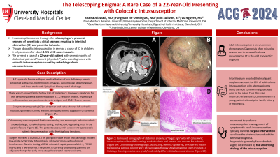
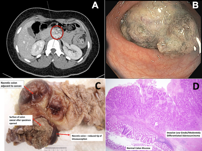
Case Description/Methods: A 22-year-old female with past medical history of iron deficiency anemia presented with a five-month history of nausea, periumbilical abdominal pain, and loose stools with “jelly-like” bloody rectal discharge. There was no known family history (FH) of malignancy. Labs were significant for iron deficiency anemia with hemoglobin 8.1. C-reactive protein, erythrocyte sedimentation rate, carcinoembryonic antigen, and CA 19-9 were normal. Computed tomography (CT) of abdomen and pelvis showed left colocolic intussusception with colonic wall thickening and edema suggestive of ischemia (Figure 1A). Colonoscopy was completed for tissue sampling and endoscopic reduction which showed a large, completely obstructing, and necrotic-appearing mass in the splenic flexure (Figure 1B). The patient subsequently underwent laparoscopic splenic flexure resection with diverting loop ileostomy. Surgery revealed a 5.0x4.5x2.0 cm polypoid friable lesion and pathology showed low-grade moderately differentiated adenocarcinoma without nodal involvement. Genetic testing of DNA mismatch repair proteins MLH-1, PMS-2, MSH-2 and 6 were normal. The patient is currently undergoing planning for adjuvant therapy for early onset stage II colorectal adenocarcinoma.
Discussion: Adult intussusception is uncommon. Diagnosis is often missed or delayed due to nonspecific clinical presentations. CT is the gold standard for diagnosis. Prior literature reported that malignant neoplasms account for 66% of adult colonic intussusception, with adenocarcinoma being the most common malignant lead point in the colon. Thus, this is an important differential to consider even in a young patient without FH of malignancy. In contrast to pediatric intussusception, management of symptomatic adult intussusception typically involves surgical intervention to relieve the obstruction and aid the definitive diagnosis. Prognosis is generally favorable but largely determined by the underlying etiology of the intussusception.

Disclosures:
Shaina Ailawadi, MD1, Fangyuan Jin-Dominguez, MD1, Erin Sullivan, BS1, Joseph Willis, MD1, Vu Q. Nguyen, MD, MS2. P2026 - The Telescoping Enigma: A Rare Case of a 22-Year-Old Presenting With Colocolonic Intussusception, ACG 2024 Annual Scientific Meeting Abstracts. Philadelphia, PA: American College of Gastroenterology.
1University Hospitals Cleveland Medical Center, Case Western Reserve University, Cleveland, OH; 2Digestive Health Institute, University Hospitals Cleveland Medical Center, Cleveland, OH
Introduction: Intussusception occurs through the telescoping of a proximal segment of bowel into a distal segment resulting in intestinal obstruction (IO) and potential ischemia. Though idiopathic intussusception is seen as a cause of IO in children, it only accounts for about 1-5% of IO cases in adults. We present a case of a 22-year-old patient with several months of abdominal pain and “currant-jelly stools”, who was diagnosed with colocolic intussusception caused by underlying colonic adenocarcinoma.
Case Description/Methods: A 22-year-old female with past medical history of iron deficiency anemia presented with a five-month history of nausea, periumbilical abdominal pain, and loose stools with “jelly-like” bloody rectal discharge. There was no known family history (FH) of malignancy. Labs were significant for iron deficiency anemia with hemoglobin 8.1. C-reactive protein, erythrocyte sedimentation rate, carcinoembryonic antigen, and CA 19-9 were normal. Computed tomography (CT) of abdomen and pelvis showed left colocolic intussusception with colonic wall thickening and edema suggestive of ischemia (Figure 1A). Colonoscopy was completed for tissue sampling and endoscopic reduction which showed a large, completely obstructing, and necrotic-appearing mass in the splenic flexure (Figure 1B). The patient subsequently underwent laparoscopic splenic flexure resection with diverting loop ileostomy. Surgery revealed a 5.0x4.5x2.0 cm polypoid friable lesion and pathology showed low-grade moderately differentiated adenocarcinoma without nodal involvement. Genetic testing of DNA mismatch repair proteins MLH-1, PMS-2, MSH-2 and 6 were normal. The patient is currently undergoing planning for adjuvant therapy for early onset stage II colorectal adenocarcinoma.
Discussion: Adult intussusception is uncommon. Diagnosis is often missed or delayed due to nonspecific clinical presentations. CT is the gold standard for diagnosis. Prior literature reported that malignant neoplasms account for 66% of adult colonic intussusception, with adenocarcinoma being the most common malignant lead point in the colon. Thus, this is an important differential to consider even in a young patient without FH of malignancy. In contrast to pediatric intussusception, management of symptomatic adult intussusception typically involves surgical intervention to relieve the obstruction and aid the definitive diagnosis. Prognosis is generally favorable but largely determined by the underlying etiology of the intussusception.

Figure: Computed tomography of abdomen showing a “target sign” with left colocolonic intussusception with wall thickening, marked colonic wall edema, and concern for ischemia (Figure 1A). Colonoscopy showing large, obstructing, necrotic-appearing, protuberant mass in the proximal sigmoid colon (Figure 1B). Colon resection specimen post-initial fixation (Figure 1C). Invasive moderately differentiated adenocarcinoma of colon (H&E stain, 20x magnification) (Figure 1D).
Disclosures:
Shaina Ailawadi indicated no relevant financial relationships.
Fangyuan Jin-Dominguez indicated no relevant financial relationships.
Erin Sullivan indicated no relevant financial relationships.
Joseph Willis indicated no relevant financial relationships.
Vu Nguyen: AbbVie – Speakers Bureau. Eli Lilly – Speakers Bureau.
Shaina Ailawadi, MD1, Fangyuan Jin-Dominguez, MD1, Erin Sullivan, BS1, Joseph Willis, MD1, Vu Q. Nguyen, MD, MS2. P2026 - The Telescoping Enigma: A Rare Case of a 22-Year-Old Presenting With Colocolonic Intussusception, ACG 2024 Annual Scientific Meeting Abstracts. Philadelphia, PA: American College of Gastroenterology.
