Monday Poster Session
Category: Colon
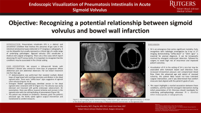
P2047 - Endoscopic Visualization of Pneumatosis Intestinalis in Acute Sigmoid Volvulus
Monday, October 28, 2024
10:30 AM - 4:00 PM ET
Location: Exhibit Hall E

Has Audio
.jpg)
Arvind Bussetty, MD
Robert Wood Johnson Medical School, Rutgers University, NJ
Presenting Author(s)
Arvind Bussetty, MD, Ping He, MD, PhD, Anish V. Patel, MD
Robert Wood Johnson Medical School, Rutgers University, New Brunswick, NJ
Introduction: Pneumatosis intestinalis (PI) is a distinct and uncommon condition that involves the presence of gas cysts in the intestinal extraluminal space detected in CT imaging or radiography. It can be idiopathic but usually represents a clinical sign of a wide range of underlying pathologies. Sigmoid volvulus (SV) constitutes a medical and surgical emergency. There are a few cases which report observing SV and PI concurrently. It is important to recognize that the conditions may be associated in the clinical setting.
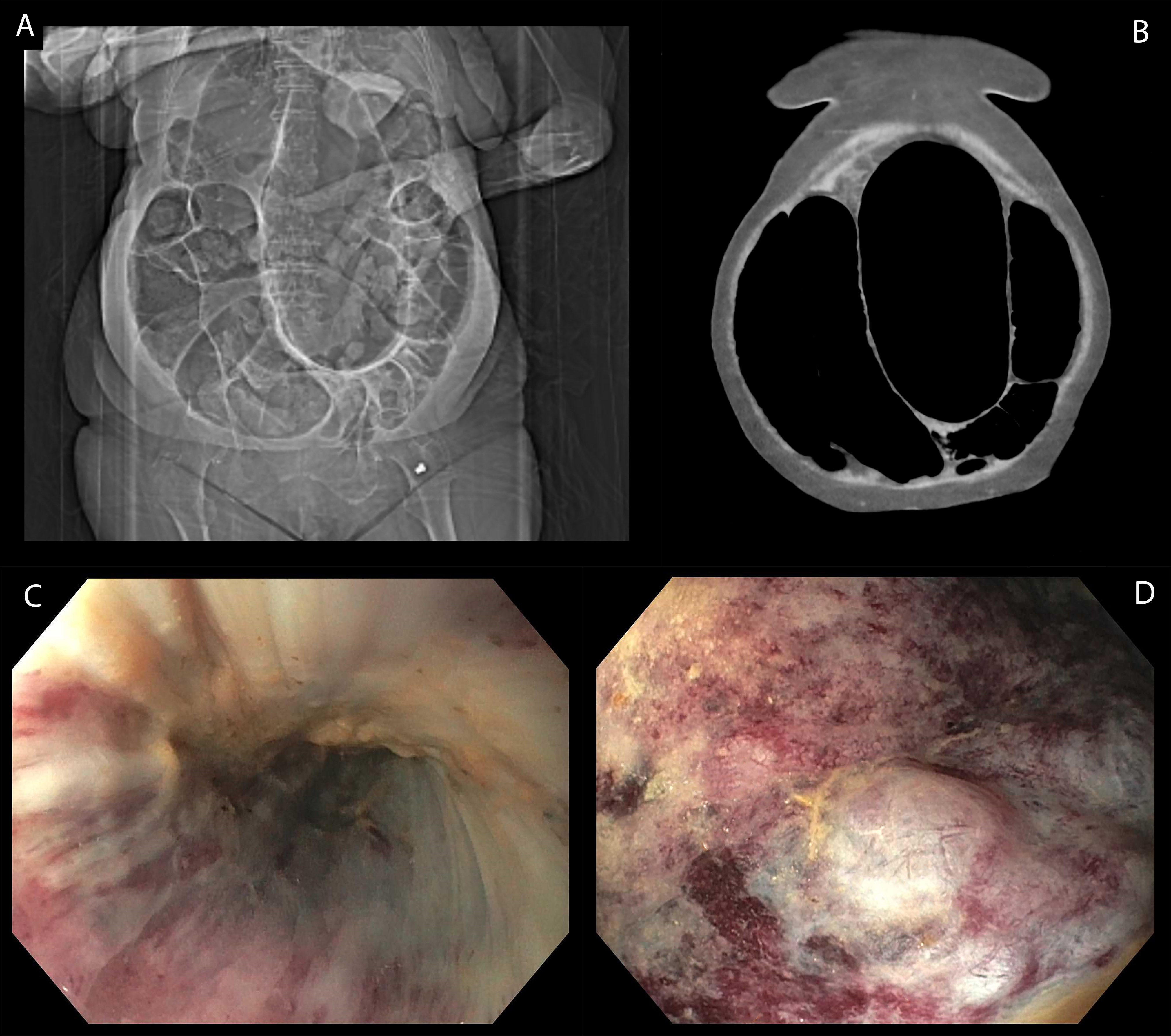
Case Description/Methods: We present a 100-year-old female with Alzheimer’s disease who arrived for three days of progressive diffuse abdominal pain and abdominal distension. Her last bowel movement was four days ago. CT abdomen/pelvis was performed that revealed multiple dilated loops of large bowel with swirling mesentery and dilation in the distal sigmoid colon. There were “coffee bean” signs suggestive of sigmoid colon volvulus (Figures 1A, B). An emergent flexible sigmoidoscopy identified torsion in the distal sigmoid colon consistent with SV (Figure 1C). The volvulus was easily detorsed and traversed with gentle endoscopic advancement. On endoscopic evaluation, there was diffuse mucosal ischemia and necrosis in the sigmoid colon with the striking finding of pneumatosis (Figure 1D). The patient was initiated on antibiotics. However, given the patient’s advanced age, the family opted against surgical intervention and transitioned to comfort measures.
Discussion: SV is an emergency that carries significant mortality. Early recognition with radiologic investigation by X-ray or CT imaging demonstrating “coffee-bean” or “whirl signs” is essential for prompt diagnosis. It can be temporarily resolved by emergent endoscopic detorsion, followed by surgery to avoid high risk of recurrence and improved patient outcomes. Visualization of PI in the setting of SV is rare but may be associated with fulminant bowel wall infarction from increased intraluminal pressure and compromised blood flow. Given the advanced age and extent of mucosal ischemia, this patient likely would not have tolerated surgical intervention, and it was determined that comfort measures best aligned with the patient’s goals of care. This report highlights a morbid association between these conditions, and the need for emergent intervention during initial presentation of SV. Clinicians should investigate for underlying pathophysiology whenever PI is identified either endoscopically or radiographically

Disclosures:
Arvind Bussetty, MD, Ping He, MD, PhD, Anish V. Patel, MD. P2047 - Endoscopic Visualization of Pneumatosis Intestinalis in Acute Sigmoid Volvulus, ACG 2024 Annual Scientific Meeting Abstracts. Philadelphia, PA: American College of Gastroenterology.
Robert Wood Johnson Medical School, Rutgers University, New Brunswick, NJ
Introduction: Pneumatosis intestinalis (PI) is a distinct and uncommon condition that involves the presence of gas cysts in the intestinal extraluminal space detected in CT imaging or radiography. It can be idiopathic but usually represents a clinical sign of a wide range of underlying pathologies. Sigmoid volvulus (SV) constitutes a medical and surgical emergency. There are a few cases which report observing SV and PI concurrently. It is important to recognize that the conditions may be associated in the clinical setting.
Case Description/Methods: We present a 100-year-old female with Alzheimer’s disease who arrived for three days of progressive diffuse abdominal pain and abdominal distension. Her last bowel movement was four days ago. CT abdomen/pelvis was performed that revealed multiple dilated loops of large bowel with swirling mesentery and dilation in the distal sigmoid colon. There were “coffee bean” signs suggestive of sigmoid colon volvulus (Figures 1A, B). An emergent flexible sigmoidoscopy identified torsion in the distal sigmoid colon consistent with SV (Figure 1C). The volvulus was easily detorsed and traversed with gentle endoscopic advancement. On endoscopic evaluation, there was diffuse mucosal ischemia and necrosis in the sigmoid colon with the striking finding of pneumatosis (Figure 1D). The patient was initiated on antibiotics. However, given the patient’s advanced age, the family opted against surgical intervention and transitioned to comfort measures.
Discussion: SV is an emergency that carries significant mortality. Early recognition with radiologic investigation by X-ray or CT imaging demonstrating “coffee-bean” or “whirl signs” is essential for prompt diagnosis. It can be temporarily resolved by emergent endoscopic detorsion, followed by surgery to avoid high risk of recurrence and improved patient outcomes. Visualization of PI in the setting of SV is rare but may be associated with fulminant bowel wall infarction from increased intraluminal pressure and compromised blood flow. Given the advanced age and extent of mucosal ischemia, this patient likely would not have tolerated surgical intervention, and it was determined that comfort measures best aligned with the patient’s goals of care. This report highlights a morbid association between these conditions, and the need for emergent intervention during initial presentation of SV. Clinicians should investigate for underlying pathophysiology whenever PI is identified either endoscopically or radiographically

Figure: Figure 1. A: Abdominal X-Ray revealing significant colonic distension. B: CT Abdomen/Pelvic demonstrating colonic distension concerning for volvulus. C and D: Pneumatosis Intestinalis findings seen on colonoscopy.
Disclosures:
Arvind Bussetty indicated no relevant financial relationships.
Ping He indicated no relevant financial relationships.
Anish Patel indicated no relevant financial relationships.
Arvind Bussetty, MD, Ping He, MD, PhD, Anish V. Patel, MD. P2047 - Endoscopic Visualization of Pneumatosis Intestinalis in Acute Sigmoid Volvulus, ACG 2024 Annual Scientific Meeting Abstracts. Philadelphia, PA: American College of Gastroenterology.
