Monday Poster Session
Category: Colon
P2054 - Intestinal Myiasis: A Seldom-Detected Cause of Chronic Upper GI Symptoms
Monday, October 28, 2024
10:30 AM - 4:00 PM ET
Location: Exhibit Hall E

Has Audio

Faris Shweikeh, MD
Cleveland Clinic Akron General
Akron, OH
Presenting Author(s)
Faris Shweikeh, MD1, Colin Fricker, BA2, Neha Sharma, MD3, Naga Venkata Rama Krishna Vura, MD4, Raja Chandra Chakinala, MD5, Mohamad Mouchli, MD6
1Cleveland Clinic Akron General, Akron, OH; 2Northeast Ohio Medical University, Rootstown, OH; 3University of Texas Health Science Center, San Antonio, TX; 4University of Texas Health San Antonio, San Antonio, TX; 5Guthrie Robert Packer Hospital, Sayre, PA; 6Cleveland Clinic, Cleveland, OH
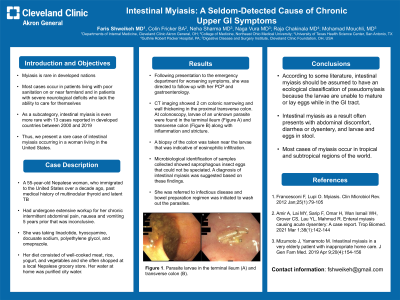
Introduction: Myiasis is rare in developed nations with most cases occurring in patients living with poor sanitation on or near farmland and in patients with severe neurological deficits who lack the ability to care for themselves. As a subcategory, intestinal myiasis is even more rare with 13 cases reported in developed countries between 2000 and 2019. Thus, we present a rare case of intestinal myiasis occurring in a woman living in the United States.
Case Description/Methods: A 55-year-old Nepalese woman, who immigrated to the United States over a decade ago, with a past medical history of multinodular thyroid and latent TB had undergone extensive workup for her chronic intermittent abdominal pain, nausea and vomiting 5 years prior that was inconclusive. She was taking linaclotide, hyoscyamine, docusate sodium, polyethylene glycol, and omeprazole. Her diet consisted of well-cooked meat, rice, yogurt, and vegetables and she often shopped at a local Nepalese grocery store. Her water at home was purified city water.
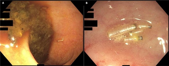
Following presentation to the emergency department for worsening symptoms, she was directed to follow-up with her PCP and gastroenterology. CT imaging showed 2 cm colonic narrowing and wall thickening in the proximal transverse colon. At colonoscopy, larvae of an unknown parasite were found in the terminal ileum (Figure A) and transverse colon (Figure B) along with inflammation and stricture. A biopsy of the colon was taken near the larvae that was indicative of eosinophilic infiltration. Microbiological identification of samples collected showed saprophagous insect eggs that could not be speciated. A diagnosis of intestinal myiasis was suggested based on these findings. She was referred to infectious disease and bowel preparation regimen was initiated to wash out the parasites.
Discussion: According to some literature, intestinal myiasis should be assumed to have an ecological classification of pseudomyiasis because the larvae are unable to mature or lay eggs while in the GI tract. Intestinal myiasis as a result often presents with abdominal discomfort, diarrhea or dysentery, and larvae and eggs in stool. Most cases of myiasis occur in tropical and subtropical regions of the world.

Disclosures:
Faris Shweikeh, MD1, Colin Fricker, BA2, Neha Sharma, MD3, Naga Venkata Rama Krishna Vura, MD4, Raja Chandra Chakinala, MD5, Mohamad Mouchli, MD6. P2054 - Intestinal Myiasis: A Seldom-Detected Cause of Chronic Upper GI Symptoms, ACG 2024 Annual Scientific Meeting Abstracts. Philadelphia, PA: American College of Gastroenterology.
1Cleveland Clinic Akron General, Akron, OH; 2Northeast Ohio Medical University, Rootstown, OH; 3University of Texas Health Science Center, San Antonio, TX; 4University of Texas Health San Antonio, San Antonio, TX; 5Guthrie Robert Packer Hospital, Sayre, PA; 6Cleveland Clinic, Cleveland, OH
Introduction: Myiasis is rare in developed nations with most cases occurring in patients living with poor sanitation on or near farmland and in patients with severe neurological deficits who lack the ability to care for themselves. As a subcategory, intestinal myiasis is even more rare with 13 cases reported in developed countries between 2000 and 2019. Thus, we present a rare case of intestinal myiasis occurring in a woman living in the United States.
Case Description/Methods: A 55-year-old Nepalese woman, who immigrated to the United States over a decade ago, with a past medical history of multinodular thyroid and latent TB had undergone extensive workup for her chronic intermittent abdominal pain, nausea and vomiting 5 years prior that was inconclusive. She was taking linaclotide, hyoscyamine, docusate sodium, polyethylene glycol, and omeprazole. Her diet consisted of well-cooked meat, rice, yogurt, and vegetables and she often shopped at a local Nepalese grocery store. Her water at home was purified city water.
Following presentation to the emergency department for worsening symptoms, she was directed to follow-up with her PCP and gastroenterology. CT imaging showed 2 cm colonic narrowing and wall thickening in the proximal transverse colon. At colonoscopy, larvae of an unknown parasite were found in the terminal ileum (Figure A) and transverse colon (Figure B) along with inflammation and stricture. A biopsy of the colon was taken near the larvae that was indicative of eosinophilic infiltration. Microbiological identification of samples collected showed saprophagous insect eggs that could not be speciated. A diagnosis of intestinal myiasis was suggested based on these findings. She was referred to infectious disease and bowel preparation regimen was initiated to wash out the parasites.
Discussion: According to some literature, intestinal myiasis should be assumed to have an ecological classification of pseudomyiasis because the larvae are unable to mature or lay eggs while in the GI tract. Intestinal myiasis as a result often presents with abdominal discomfort, diarrhea or dysentery, and larvae and eggs in stool. Most cases of myiasis occur in tropical and subtropical regions of the world.

Figure: Parasite larvae in the terminal ileum (A) and transverse colon (B).
Disclosures:
Faris Shweikeh indicated no relevant financial relationships.
Colin Fricker indicated no relevant financial relationships.
Neha Sharma indicated no relevant financial relationships.
Naga Venkata Rama Krishna Vura indicated no relevant financial relationships.
Raja Chandra Chakinala indicated no relevant financial relationships.
Mohamad Mouchli indicated no relevant financial relationships.
Faris Shweikeh, MD1, Colin Fricker, BA2, Neha Sharma, MD3, Naga Venkata Rama Krishna Vura, MD4, Raja Chandra Chakinala, MD5, Mohamad Mouchli, MD6. P2054 - Intestinal Myiasis: A Seldom-Detected Cause of Chronic Upper GI Symptoms, ACG 2024 Annual Scientific Meeting Abstracts. Philadelphia, PA: American College of Gastroenterology.
