Monday Poster Session
Category: Colon
P2094 - Squamous Cell Carcinoma of the Rectum: A Review of Endoscopic Features and Case Report
Monday, October 28, 2024
10:30 AM - 4:00 PM ET
Location: Exhibit Hall E

Has Audio

Rohan Rau, DO
Albert Einstein Medical Center
Philadelphia, PA
Presenting Author(s)
Rohan Rau, DO1, Edward J. Marines, DO2, Matthew Chan, DO3, Michael L. Davis, DO1
1Albert Einstein Medical Center, Philadelphia, PA; 2Jefferson Einstein Hospital, Philadelphia, PA; 3Einstein Healthcare Network, East Norriton, PA
Introduction: Squamous cell carcinoma (SCC) of the lower gastrointestinal tract is most commonly associated with anal cancer. Here we present a case of rectal SCC (RSCC). A review of the literature was conducted for endoscopic features of RSCC.
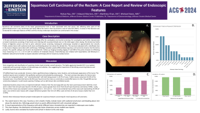
Case Description/Methods: A 54-year-old female presented to the gastroenterology office for constipation and routine screening. Subsequent colonoscopy revealed a 2cm smooth, friable, nodular lesion with scattered small ulcers and bleeding located about 2 cm above the dentate line. Biopsies were taken with cold forceps and pathology revealed a poorly differentiated squamous cell carcinoma invasive into the lamina propria. Chemotherapy and radiation reduced tumor burden as repeat evaluation with biopsy did not reveal any residual cancer.
Discussion: Early recognition and classification of suspicious lesions leads to better overall outcomes. The highly aggressive basaloid SCC in our patient responded well to prompt initiation of chemotherapy and radiation, thus suggesting the importance of identifying concerning features and commencing proper workup without delay.
A PubMed Search was conducted. Inclusion criteria specified primary malignancy, tumor location, and endoscopic appearance of the tumor. The resultant reports were included in the qualitative synthesis and evaluated by endoscopists.
The most common descriptors for lesions were found to be “polypoid/nodular” and “ulcerated/eroded” with both being present in 30.7% of cases. Friable masses comprised 19.23% of the reviewed cases. Circumferential lesions (26.9%) were found more than twice as commonly as sessile/flat lesions (11.5%). Though NBI was less commonly employed, it could reveal suspicious findings such as dark brown dots and dilated vessels.
Anatomical location proves to be an important landmark. Distance from the anal verge (AV) was the most commonly used measurement. Both the 1-5cm and 5.1-10cm from AV each were found in 19.23% of cases. Lesions greater than 10 cm from the AV were found less commonly (7.69%).
Lastly, the size of the masses was evaluated. Lesions ranging from 1- 3cm and 3.1 - 5cm in size contained many of the cases each representing 19.23% of cases. Less common were masses with a largest dimension greater than 5cm (15.38%). Least common of all were masses smaller than 1cm (7.69%).
Overall, these endoscopic features prepare endoscopists for suspicious features concerning for rectal squamous cell carcinoma. Early recognition expedites treatment and hopefully improves outcomes as well.

Disclosures:
Rohan Rau, DO1, Edward J. Marines, DO2, Matthew Chan, DO3, Michael L. Davis, DO1. P2094 - Squamous Cell Carcinoma of the Rectum: A Review of Endoscopic Features and Case Report, ACG 2024 Annual Scientific Meeting Abstracts. Philadelphia, PA: American College of Gastroenterology.
1Albert Einstein Medical Center, Philadelphia, PA; 2Jefferson Einstein Hospital, Philadelphia, PA; 3Einstein Healthcare Network, East Norriton, PA
Introduction: Squamous cell carcinoma (SCC) of the lower gastrointestinal tract is most commonly associated with anal cancer. Here we present a case of rectal SCC (RSCC). A review of the literature was conducted for endoscopic features of RSCC.
Case Description/Methods: A 54-year-old female presented to the gastroenterology office for constipation and routine screening. Subsequent colonoscopy revealed a 2cm smooth, friable, nodular lesion with scattered small ulcers and bleeding located about 2 cm above the dentate line. Biopsies were taken with cold forceps and pathology revealed a poorly differentiated squamous cell carcinoma invasive into the lamina propria. Chemotherapy and radiation reduced tumor burden as repeat evaluation with biopsy did not reveal any residual cancer.
Discussion: Early recognition and classification of suspicious lesions leads to better overall outcomes. The highly aggressive basaloid SCC in our patient responded well to prompt initiation of chemotherapy and radiation, thus suggesting the importance of identifying concerning features and commencing proper workup without delay.
A PubMed Search was conducted. Inclusion criteria specified primary malignancy, tumor location, and endoscopic appearance of the tumor. The resultant reports were included in the qualitative synthesis and evaluated by endoscopists.
The most common descriptors for lesions were found to be “polypoid/nodular” and “ulcerated/eroded” with both being present in 30.7% of cases. Friable masses comprised 19.23% of the reviewed cases. Circumferential lesions (26.9%) were found more than twice as commonly as sessile/flat lesions (11.5%). Though NBI was less commonly employed, it could reveal suspicious findings such as dark brown dots and dilated vessels.
Anatomical location proves to be an important landmark. Distance from the anal verge (AV) was the most commonly used measurement. Both the 1-5cm and 5.1-10cm from AV each were found in 19.23% of cases. Lesions greater than 10 cm from the AV were found less commonly (7.69%).
Lastly, the size of the masses was evaluated. Lesions ranging from 1- 3cm and 3.1 - 5cm in size contained many of the cases each representing 19.23% of cases. Less common were masses with a largest dimension greater than 5cm (15.38%). Least common of all were masses smaller than 1cm (7.69%).
Overall, these endoscopic features prepare endoscopists for suspicious features concerning for rectal squamous cell carcinoma. Early recognition expedites treatment and hopefully improves outcomes as well.

Figure: Figure 1. Colonoscopy of the rectum showing a 2cm smooth, friable, nodular lesion with scattered small ulcers and bleeding.
Figure 2: Chart of endoscopic features of rectal squamous cell carcinoma upon review of the literature.
Figure 3. Chart detailing location of rectal SCC masses in the literature.
Figure 4. Chart illustrating the size categories of masses that were identified as rectal squamous cell carcinoma.
Figure 2: Chart of endoscopic features of rectal squamous cell carcinoma upon review of the literature.
Figure 3. Chart detailing location of rectal SCC masses in the literature.
Figure 4. Chart illustrating the size categories of masses that were identified as rectal squamous cell carcinoma.
Disclosures:
Rohan Rau indicated no relevant financial relationships.
Edward Marines indicated no relevant financial relationships.
Matthew Chan indicated no relevant financial relationships.
Michael Davis indicated no relevant financial relationships.
Rohan Rau, DO1, Edward J. Marines, DO2, Matthew Chan, DO3, Michael L. Davis, DO1. P2094 - Squamous Cell Carcinoma of the Rectum: A Review of Endoscopic Features and Case Report, ACG 2024 Annual Scientific Meeting Abstracts. Philadelphia, PA: American College of Gastroenterology.
