Monday Poster Session
Category: Endoscopy Video Forum
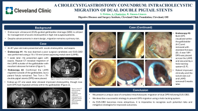
P2177 - A Cholecystgastrostomy Conundrum: Intracholecystic Migration of Dual Double Pigtail Stents
Monday, October 28, 2024
10:30 AM - 4:00 PM ET
Location: Exhibit Hall E

Has Audio

Stephen Firkins, MD
Cleveland Clinic
Cleveland, OH
Presenting Author(s)
Stephen Firkins, MD1, Arjun Chatterjee, MD2, Prabhleen Chahal, MD2, C Roberto Simon-Linares, MD1
1Cleveland Clinic, Cleveland, OH; 2Cleveland Clinic Foundation, Cleveland, OH
Introduction: The American Gastroenterological Association suggests endoscopic ultrasound (EUS)-guided gallbladder drainage (GBD) for management of acute cholecystitis in high-risk surgical patients. Despite the advantageous design of lumen apposing metal stents (LEMS), migration remains a primary risk of EUS-GBD.
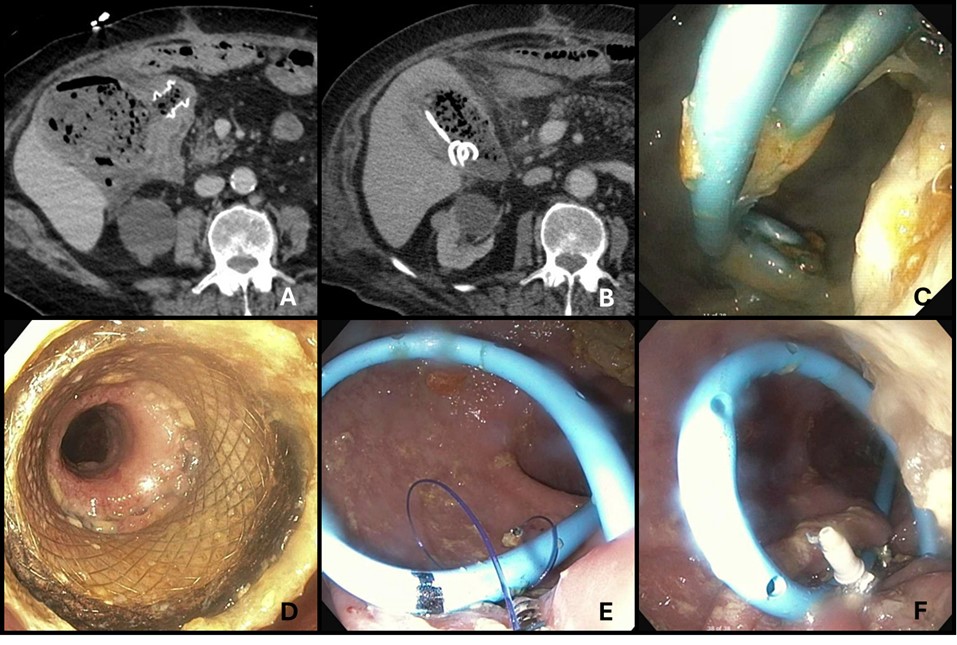
Case Description/Methods: An 87-year-old man with a complex surgical history including toxic colitis status post subtotal colectomy with end ileostomy and recurrent small bowel obstructions presented with sepsis. Computed tomography (CT) revealed marked gallbladder distension and pericholecystic fluid consistent with acute cholecystitis. He was deemed to be a poor surgical candidate and thus EUS-GBD was pursued. EUS revealed a distended gallbladder with extensive sludge and small stones, and a 15mm x 10mm LAMS was successfully placed for drainage. He was discharged several days later on oral antibiotics, however presented 1 week later with recurrent sepsis. A repeat CT scan revealed displacement of the LAMS outside of the gallbladder and interval development of a 6.5cm x 7.3cm pericholecystic abscess (Figure 1A). Subsequent endoscopy confirmed the LAMS migrated outside of the gallbladder but within an intact fistula allowing access to the gallbladder, and two 7cm x 7Fr double pigtail stents (DPS) were placed across the LAMS. Follow-up CT one week later showed improved gallbladder, though now both DPS had migrated entirely within the gallbladder (Figure 1B). A third endoscopy was performed, finding both DPS within the gallbladder (Figure 1C). The migrated DPS were removed and a new 10Fr DPS was placed across the LAMS for drainage and secured by a helix tacking system (Figure 1D-F). The patient improved clinically over the subsequent weeks. A final endoscopy was performed 6 weeks later, during which the helix tacking suture was cut and the LAMS removed.
Discussion: Endoscopic drainage of acute cholecystitis is increasingly utilized in poor operative candidates. While the double-flange design of LAMS is intended to reduce migration risk, stent migration in EUS-GBD still occurs in roughly 3% of cases. We present a unique case of LAMS migration followed by complete intracholecystic migration of dual DPS after EUS-GBD and describe a successful strategy to prevent DPS migration using a helix tacking system. As EUS-GBD becomes more ubiquitous, it is imperative to recognize such potential risks and mitigation strategies for improved outcomes.

Disclosures:
Stephen Firkins, MD1, Arjun Chatterjee, MD2, Prabhleen Chahal, MD2, C Roberto Simon-Linares, MD1. P2177 - A Cholecystgastrostomy Conundrum: Intracholecystic Migration of Dual Double Pigtail Stents, ACG 2024 Annual Scientific Meeting Abstracts. Philadelphia, PA: American College of Gastroenterology.
1Cleveland Clinic, Cleveland, OH; 2Cleveland Clinic Foundation, Cleveland, OH
Introduction: The American Gastroenterological Association suggests endoscopic ultrasound (EUS)-guided gallbladder drainage (GBD) for management of acute cholecystitis in high-risk surgical patients. Despite the advantageous design of lumen apposing metal stents (LEMS), migration remains a primary risk of EUS-GBD.
Case Description/Methods: An 87-year-old man with a complex surgical history including toxic colitis status post subtotal colectomy with end ileostomy and recurrent small bowel obstructions presented with sepsis. Computed tomography (CT) revealed marked gallbladder distension and pericholecystic fluid consistent with acute cholecystitis. He was deemed to be a poor surgical candidate and thus EUS-GBD was pursued. EUS revealed a distended gallbladder with extensive sludge and small stones, and a 15mm x 10mm LAMS was successfully placed for drainage. He was discharged several days later on oral antibiotics, however presented 1 week later with recurrent sepsis. A repeat CT scan revealed displacement of the LAMS outside of the gallbladder and interval development of a 6.5cm x 7.3cm pericholecystic abscess (Figure 1A). Subsequent endoscopy confirmed the LAMS migrated outside of the gallbladder but within an intact fistula allowing access to the gallbladder, and two 7cm x 7Fr double pigtail stents (DPS) were placed across the LAMS. Follow-up CT one week later showed improved gallbladder, though now both DPS had migrated entirely within the gallbladder (Figure 1B). A third endoscopy was performed, finding both DPS within the gallbladder (Figure 1C). The migrated DPS were removed and a new 10Fr DPS was placed across the LAMS for drainage and secured by a helix tacking system (Figure 1D-F). The patient improved clinically over the subsequent weeks. A final endoscopy was performed 6 weeks later, during which the helix tacking suture was cut and the LAMS removed.
Discussion: Endoscopic drainage of acute cholecystitis is increasingly utilized in poor operative candidates. While the double-flange design of LAMS is intended to reduce migration risk, stent migration in EUS-GBD still occurs in roughly 3% of cases. We present a unique case of LAMS migration followed by complete intracholecystic migration of dual DPS after EUS-GBD and describe a successful strategy to prevent DPS migration using a helix tacking system. As EUS-GBD becomes more ubiquitous, it is imperative to recognize such potential risks and mitigation strategies for improved outcomes.

Figure: Figure 1
A: CT scan showing LAMS migration outside of the gallbladder lumen with resultant pericholecystic abscess
B: CT scan showing both DPS visualized within the gallbladder
C: Endoscopic view of both DPS entirely within the gallbladder
D: Clear cholecystgastrostomy tract after DPS removal and gallbladder lavage
E-F: Helix tacking system used to secure new DPS to the gastric wall
Disclosures:
Stephen Firkins indicated no relevant financial relationships.
Arjun Chatterjee indicated no relevant financial relationships.
Prabhleen Chahal: Boston Scientific – Advisor or Review Panel Member.
C Roberto Simon-Linares indicated no relevant financial relationships.
Stephen Firkins, MD1, Arjun Chatterjee, MD2, Prabhleen Chahal, MD2, C Roberto Simon-Linares, MD1. P2177 - A Cholecystgastrostomy Conundrum: Intracholecystic Migration of Dual Double Pigtail Stents, ACG 2024 Annual Scientific Meeting Abstracts. Philadelphia, PA: American College of Gastroenterology.

