Monday Poster Session
Category: Esophagus
P2250 - A Rare Case of Complete Esophageal Obstruction Following Recurrent Banding of Gastroesophageal Varices
Monday, October 28, 2024
10:30 AM - 4:00 PM ET
Location: Exhibit Hall E

Has Audio

Trisha Andrews, MD
Riverside Medical Center
Kankakee, IL
Presenting Author(s)
Trisha Andrews, MD, Matthew Sutherland, DO, Kyle Hamann, DO, Nancy Mayer, DO, Brian Sasso, DO
Riverside Medical Center, Kankakee, IL
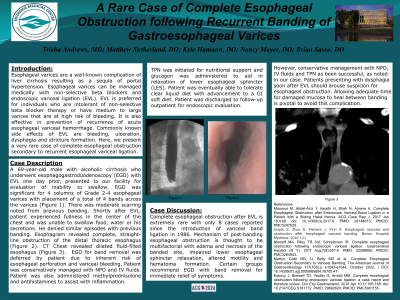
Introduction: Esophageal varices are a well-known complication of liver cirrhosis resulting as a sequela of portal hypertension. Esophageal varices can be managed medically with non-selective beta blockers and endoscopic variceal ligation (EVL). EVL is preferred for individuals who are intolerant of non-selective beta blocker therapy or have medium to large varices that are at a higher risk of bleeding. It is also effective in prevention of recurrence of acute esophageal variceal hemorrhage. Common side effects of EVL are bleeding, ulceration, dysphagia and stricture formation. We present a rare case of complete esophageal obstruction secondary to recurrent EVL.
Case Description/Methods: A 69-year-old male with alcoholic cirrhosis who underwent EGD with EVL one day prior presented with an inability to swallow. EGD showed moderate scarring from prior banding and 4 columns of Grade 2-4 esophageal varices (Fig. 1) with placement of 4 bands across the varices. Shortly after EVL, patient experienced fullness in the center of the chest and was unable to swallow food, water or his secretions. He denied similar episodes with prior banding. Esophagram demonstrated complete, straight-line obstruction of the distal thoracic esophagus (Fig. 2). CT chest revealed dilated fluid-filled esophagus (Fig. 3). EGD for band removal was deferred by patient due to risk of esophageal perforation and variceal bleeding. Patient was conservatively managed with NPO and IV fluids. Methylprednisolone and antihistamines were administered to reduce inflammation.TPN was initiated for nutritional support and glucagon was administered to aid in relaxation of lower esophageal sphincter (LES). Patient was eventually able to tolerate clear liquid diet with advancement to GI soft diet. Patient was discharged to follow-up outpatient for endoscopic evaluation.
Discussion: Complete esophageal obstruction after EVL is extremely rare with only 8 cases reported since the introduction of EVL in 1986. Mechanism of post-banding esophageal obstruction is thought to be multifactorial with edema and necrosis of the banded site, impaired LES relaxation, altered motility and hematoma formation. EGD with band removal can bring immediate relief of symptoms. However, conservative management with NPO, IV fluids and TPN has been successful, as noted in our case. Patients presenting with dysphagia soon after EVL should arouse suspicion for esophageal obstruction. Allowing adequate time for damaged mucosa to heal between banding is pivotal to avoid this complication.

Disclosures:
Trisha Andrews, MD, Matthew Sutherland, DO, Kyle Hamann, DO, Nancy Mayer, DO, Brian Sasso, DO. P2250 - A Rare Case of Complete Esophageal Obstruction Following Recurrent Banding of Gastroesophageal Varices, ACG 2024 Annual Scientific Meeting Abstracts. Philadelphia, PA: American College of Gastroenterology.
Riverside Medical Center, Kankakee, IL
Introduction: Esophageal varices are a well-known complication of liver cirrhosis resulting as a sequela of portal hypertension. Esophageal varices can be managed medically with non-selective beta blockers and endoscopic variceal ligation (EVL). EVL is preferred for individuals who are intolerant of non-selective beta blocker therapy or have medium to large varices that are at a higher risk of bleeding. It is also effective in prevention of recurrence of acute esophageal variceal hemorrhage. Common side effects of EVL are bleeding, ulceration, dysphagia and stricture formation. We present a rare case of complete esophageal obstruction secondary to recurrent EVL.
Case Description/Methods: A 69-year-old male with alcoholic cirrhosis who underwent EGD with EVL one day prior presented with an inability to swallow. EGD showed moderate scarring from prior banding and 4 columns of Grade 2-4 esophageal varices (Fig. 1) with placement of 4 bands across the varices. Shortly after EVL, patient experienced fullness in the center of the chest and was unable to swallow food, water or his secretions. He denied similar episodes with prior banding. Esophagram demonstrated complete, straight-line obstruction of the distal thoracic esophagus (Fig. 2). CT chest revealed dilated fluid-filled esophagus (Fig. 3). EGD for band removal was deferred by patient due to risk of esophageal perforation and variceal bleeding. Patient was conservatively managed with NPO and IV fluids. Methylprednisolone and antihistamines were administered to reduce inflammation.TPN was initiated for nutritional support and glucagon was administered to aid in relaxation of lower esophageal sphincter (LES). Patient was eventually able to tolerate clear liquid diet with advancement to GI soft diet. Patient was discharged to follow-up outpatient for endoscopic evaluation.
Discussion: Complete esophageal obstruction after EVL is extremely rare with only 8 cases reported since the introduction of EVL in 1986. Mechanism of post-banding esophageal obstruction is thought to be multifactorial with edema and necrosis of the banded site, impaired LES relaxation, altered motility and hematoma formation. EGD with band removal can bring immediate relief of symptoms. However, conservative management with NPO, IV fluids and TPN has been successful, as noted in our case. Patients presenting with dysphagia soon after EVL should arouse suspicion for esophageal obstruction. Allowing adequate time for damaged mucosa to heal between banding is pivotal to avoid this complication.

Figure: Figure 1: EGD showing 4 columns of esophageal varices
Figure 2: Esophagram demonstrating complete, straight-line obstruction of the distal thoracic esophagus
Figure 3: CT chest showing dilated, fluid-filled esophagus
Figure 2: Esophagram demonstrating complete, straight-line obstruction of the distal thoracic esophagus
Figure 3: CT chest showing dilated, fluid-filled esophagus
Disclosures:
Trisha Andrews indicated no relevant financial relationships.
Matthew Sutherland indicated no relevant financial relationships.
Kyle Hamann indicated no relevant financial relationships.
Nancy Mayer indicated no relevant financial relationships.
Brian Sasso indicated no relevant financial relationships.
Trisha Andrews, MD, Matthew Sutherland, DO, Kyle Hamann, DO, Nancy Mayer, DO, Brian Sasso, DO. P2250 - A Rare Case of Complete Esophageal Obstruction Following Recurrent Banding of Gastroesophageal Varices, ACG 2024 Annual Scientific Meeting Abstracts. Philadelphia, PA: American College of Gastroenterology.
