Monday Poster Session
Category: Esophagus
P2261 - A Rare Presentation of Pseudoachalasia - Focusing on the Challenges and Strategies for Secondary Achalasia in Clinical Practice.
Monday, October 28, 2024
10:30 AM - 4:00 PM ET
Location: Exhibit Hall E

Has Audio

Aima Azhar, MD
Fatima Jinnah Medical University
Claymont, DE
Presenting Author(s)
Aima Azhar, MD1, Muhammad Abdul Moiz, MBBS2, Musfira Khalid, MBBS3, Faheem Javad, MBBS4, Aresha Masood Shah, MBBS5, Arsalan Hyder, MBBS6, Abdul Arham, MD7
1Fatima Jinnah Medical University, Claymont, DE; 2Services Institute of Medical Sciences, Multan, Punjab, Pakistan; 3Fatima Jinnah Medical University, Mississauga, ON, Canada; 4Al-Nafees Medical College, Mississauga, ON, Canada; 5Sindh Medical College, Karachi, Sindh, Pakistan; 6Dow Medical College, Karachi, Sindh, Pakistan; 7Baystate Medical Center, Chicopee, MA
Introduction: Pseudoachalasia refers to an achalasia-like widening of the esophagus caused by constriction in the lower part of the esophagus. With a prevalence of 2.4-4.0% of patients presenting with achalasia, Pseudoachalasia remains one of the most rarely encountered presentations in clinical settings.Due to similar clinical,radiological and endoscopic findings, distinguishing pseudoachalasia from primary achalasia poses a constant diagnostic dilemma. We present a case of a patient repeatedly being treated for aspiration pneumonia due to dysphagia who ended up having complete workup to rule out primary esophageal malignancy.
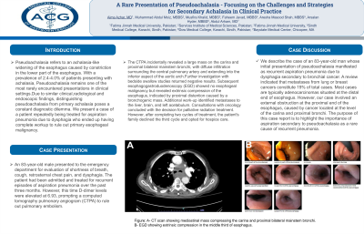
Case Description/Methods: An 83-year-old male presented to the emergency department for evaluation of shortness of breath, cough, retrosternal chest pain, and dysphagia. The patient had been admitted and treated for recurrent episodes of aspiration pneumonia over the past three months. However, this time D-dimer levels were elevated at 6.93, prompting a computed tomography pulmonary angiogram (CTPA) to rule out pulmonary embolism. The CTPA incidentally revealed a large mass on the carina and proximal bilateral mainstem bronchi, with diffuse infiltration surrounding the central pulmonary artery and extending into the inferior aspect of the aortic arch.Further investigation with bedside swallow studies returned negative results. Subsequent esophagogastroduodenoscopy (EGD) showed no esophageal malignancy but revealed extrinsic compression of the esophagus, indicated by proximal distention caused by a bronchogenic mass. Additional work-up identified metastases to the liver, brain, and left acetabulum. Consultations with oncology concluded with the decision for palliative radiation treatment. However, after completing two cycles of treatment, the patient's family declined the third cycle and opted for hospice care.
Discussion: We describe the case of an 83-year-old man whose initial presentation of pseudoachalasia manifested as recurrent aspiration pneumonia due to dysphagia secondary to bronchial cancer. A review indicated that metastases from lung or breast cancers constitute 19% of total cases. Most cases are typically adenocarcinomas situated at the distal end of esophagus. However, our case involved an external obstruction at the proximal end of the esophagus, caused by cancer located at the level of the carina and proximal bronchi.The purpose of this case report is to highlight the importance of aspiration secondary to pseudoachalasia as a rare cause of recurrent pneumonia.

Disclosures:
Aima Azhar, MD1, Muhammad Abdul Moiz, MBBS2, Musfira Khalid, MBBS3, Faheem Javad, MBBS4, Aresha Masood Shah, MBBS5, Arsalan Hyder, MBBS6, Abdul Arham, MD7. P2261 - A Rare Presentation of Pseudoachalasia - Focusing on the Challenges and Strategies for Secondary Achalasia in Clinical Practice., ACG 2024 Annual Scientific Meeting Abstracts. Philadelphia, PA: American College of Gastroenterology.
1Fatima Jinnah Medical University, Claymont, DE; 2Services Institute of Medical Sciences, Multan, Punjab, Pakistan; 3Fatima Jinnah Medical University, Mississauga, ON, Canada; 4Al-Nafees Medical College, Mississauga, ON, Canada; 5Sindh Medical College, Karachi, Sindh, Pakistan; 6Dow Medical College, Karachi, Sindh, Pakistan; 7Baystate Medical Center, Chicopee, MA
Introduction: Pseudoachalasia refers to an achalasia-like widening of the esophagus caused by constriction in the lower part of the esophagus. With a prevalence of 2.4-4.0% of patients presenting with achalasia, Pseudoachalasia remains one of the most rarely encountered presentations in clinical settings.Due to similar clinical,radiological and endoscopic findings, distinguishing pseudoachalasia from primary achalasia poses a constant diagnostic dilemma. We present a case of a patient repeatedly being treated for aspiration pneumonia due to dysphagia who ended up having complete workup to rule out primary esophageal malignancy.
Case Description/Methods: An 83-year-old male presented to the emergency department for evaluation of shortness of breath, cough, retrosternal chest pain, and dysphagia. The patient had been admitted and treated for recurrent episodes of aspiration pneumonia over the past three months. However, this time D-dimer levels were elevated at 6.93, prompting a computed tomography pulmonary angiogram (CTPA) to rule out pulmonary embolism. The CTPA incidentally revealed a large mass on the carina and proximal bilateral mainstem bronchi, with diffuse infiltration surrounding the central pulmonary artery and extending into the inferior aspect of the aortic arch.Further investigation with bedside swallow studies returned negative results. Subsequent esophagogastroduodenoscopy (EGD) showed no esophageal malignancy but revealed extrinsic compression of the esophagus, indicated by proximal distention caused by a bronchogenic mass. Additional work-up identified metastases to the liver, brain, and left acetabulum. Consultations with oncology concluded with the decision for palliative radiation treatment. However, after completing two cycles of treatment, the patient's family declined the third cycle and opted for hospice care.
Discussion: We describe the case of an 83-year-old man whose initial presentation of pseudoachalasia manifested as recurrent aspiration pneumonia due to dysphagia secondary to bronchial cancer. A review indicated that metastases from lung or breast cancers constitute 19% of total cases. Most cases are typically adenocarcinomas situated at the distal end of esophagus. However, our case involved an external obstruction at the proximal end of the esophagus, caused by cancer located at the level of the carina and proximal bronchi.The purpose of this case report is to highlight the importance of aspiration secondary to pseudoachalasia as a rare cause of recurrent pneumonia.

Figure: A- CT scan showing mediastinal mass compressing the carina and proximal bilateral mainstem bronchi.
B- EGD showing extrinsic compression in the middle third of esophagus.
B- EGD showing extrinsic compression in the middle third of esophagus.
Disclosures:
Aima Azhar indicated no relevant financial relationships.
Muhammad Abdul Moiz indicated no relevant financial relationships.
Musfira Khalid indicated no relevant financial relationships.
Faheem Javad indicated no relevant financial relationships.
Aresha Masood Shah indicated no relevant financial relationships.
Arsalan Hyder indicated no relevant financial relationships.
Abdul Arham indicated no relevant financial relationships.
Aima Azhar, MD1, Muhammad Abdul Moiz, MBBS2, Musfira Khalid, MBBS3, Faheem Javad, MBBS4, Aresha Masood Shah, MBBS5, Arsalan Hyder, MBBS6, Abdul Arham, MD7. P2261 - A Rare Presentation of Pseudoachalasia - Focusing on the Challenges and Strategies for Secondary Achalasia in Clinical Practice., ACG 2024 Annual Scientific Meeting Abstracts. Philadelphia, PA: American College of Gastroenterology.
