Monday Poster Session
Category: Esophagus
P2262 - An Extremely Rare Case of Dysphagia in Adults: Esophageal Duplication Cyst
Monday, October 28, 2024
10:30 AM - 4:00 PM ET
Location: Exhibit Hall E

Has Audio

Shreel H. Patel, MD
University of Texas Rio Grande Valley - Knapp Medical Center
Weslaco, TX
Presenting Author(s)
Shreel H. Patel, MD1, Nirav B. Patel, MS2, Dhruvkumar Patel, MBBS3, Hareesh Gundlapalli, MD4, Jawairia Memon, MD5, Valeska Balderas, MD6
1University of Texas Rio Grande Valley - Knapp Medical Center, Weslaco, TX; 2GCS Medical College, Hospital and Research Centre, Weslaco, TX; 3LSU Health Science Center, Shreveport, LA; 4University of New Mexico, San Antonio, TX; 5UTHSCSA, San Antonio, TX; 6Texas Gastroenterology Institute, McAllen, TX
Introduction: Dysphagia is subjective sensation of difficulty or abnormality of swallowing. It can be divided into oropharyngeal or esophageal dysphagia. Common causes of esophageal dysphagia are peptic stricture, carcinoma, eosinophilic esophagitis, esophageal webs and rings, or cardiovascular abnormalities leading to sensation of food not able to pass from the upper esophagus to stomach. Hence, we present a rare case of dysphagia in adults due to esophageal duplication cyst, as esophageal duplication cyst is extremely rare to see in adult population.
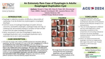
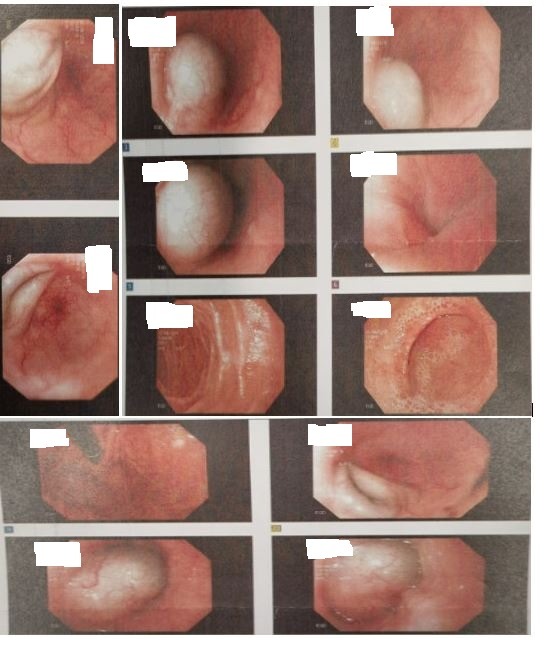
Case Description/Methods: A 37-year-old lady with history of GERD presented with chief complaint of intermittent dysphagia to solids from past 6 months associated with weight loss. She denies any nausea, pain during swallowing, melena, hematemesis, hematochezia, choking or coughing, atopic dermatitis, chest pain, palpitations or no family history of malignancy. Occasional smoker. No abnormal physical examination findings. Routine labs were normal. CT Chest with IV contrast done which showed “mediastinal mass measuring 2.7 x 3.6 x 3 cm at distal esophagus". The patient was referred to GI, EGD was done which showed large submucosal mass with no bleeding or no stigmata of recent bleeding, found in the middle third of the esophagus. The mass was non obstructing and partially circumferential, inflating and deflating on inspiration and expiration respectively and completely flattened out. It had bluish hue and appeared to be hematoma between muscularis mucosa and submucosa. EUS was done to confirm the lesion, which was hypoechoic. The patient was further referred to cardiothoracic surgeon, who performed surgical intervention, and anchovy paste like fluid was removed from the esophageal mass. The histopathology showed cyst wall consistent with the esophageal duplication cyst and fluid content showed chronic inflammation and hemosiderin-laden macrophages. Malignancy was ruled out and symptoms were improved.
Discussion: Esophageal duplication cysts are congenital anomalies of the foregut resulting from aberration of the posterior division of the embryonic foregut at 3–4-week gestation. They are mostly diagnosed in childhood and very rarely diagnosed in adults presenting as symptoms of the compression of surrounding mediastinal structures and causes dysphagia, respiratory distress, failure to thrive and retrosternal pain. Hence, our patient presented with dysphagia, and they are diagnosed by EGD, Upper EUS and CT scan. Treated surgically or with endoscopic interventions.

Disclosures:
Shreel H. Patel, MD1, Nirav B. Patel, MS2, Dhruvkumar Patel, MBBS3, Hareesh Gundlapalli, MD4, Jawairia Memon, MD5, Valeska Balderas, MD6. P2262 - An Extremely Rare Case of Dysphagia in Adults: Esophageal Duplication Cyst, ACG 2024 Annual Scientific Meeting Abstracts. Philadelphia, PA: American College of Gastroenterology.
1University of Texas Rio Grande Valley - Knapp Medical Center, Weslaco, TX; 2GCS Medical College, Hospital and Research Centre, Weslaco, TX; 3LSU Health Science Center, Shreveport, LA; 4University of New Mexico, San Antonio, TX; 5UTHSCSA, San Antonio, TX; 6Texas Gastroenterology Institute, McAllen, TX
Introduction: Dysphagia is subjective sensation of difficulty or abnormality of swallowing. It can be divided into oropharyngeal or esophageal dysphagia. Common causes of esophageal dysphagia are peptic stricture, carcinoma, eosinophilic esophagitis, esophageal webs and rings, or cardiovascular abnormalities leading to sensation of food not able to pass from the upper esophagus to stomach. Hence, we present a rare case of dysphagia in adults due to esophageal duplication cyst, as esophageal duplication cyst is extremely rare to see in adult population.
Case Description/Methods: A 37-year-old lady with history of GERD presented with chief complaint of intermittent dysphagia to solids from past 6 months associated with weight loss. She denies any nausea, pain during swallowing, melena, hematemesis, hematochezia, choking or coughing, atopic dermatitis, chest pain, palpitations or no family history of malignancy. Occasional smoker. No abnormal physical examination findings. Routine labs were normal. CT Chest with IV contrast done which showed “mediastinal mass measuring 2.7 x 3.6 x 3 cm at distal esophagus". The patient was referred to GI, EGD was done which showed large submucosal mass with no bleeding or no stigmata of recent bleeding, found in the middle third of the esophagus. The mass was non obstructing and partially circumferential, inflating and deflating on inspiration and expiration respectively and completely flattened out. It had bluish hue and appeared to be hematoma between muscularis mucosa and submucosa. EUS was done to confirm the lesion, which was hypoechoic. The patient was further referred to cardiothoracic surgeon, who performed surgical intervention, and anchovy paste like fluid was removed from the esophageal mass. The histopathology showed cyst wall consistent with the esophageal duplication cyst and fluid content showed chronic inflammation and hemosiderin-laden macrophages. Malignancy was ruled out and symptoms were improved.
Discussion: Esophageal duplication cysts are congenital anomalies of the foregut resulting from aberration of the posterior division of the embryonic foregut at 3–4-week gestation. They are mostly diagnosed in childhood and very rarely diagnosed in adults presenting as symptoms of the compression of surrounding mediastinal structures and causes dysphagia, respiratory distress, failure to thrive and retrosternal pain. Hence, our patient presented with dysphagia, and they are diagnosed by EGD, Upper EUS and CT scan. Treated surgically or with endoscopic interventions.

Figure: This image shows the pictures taken during EGD that how esophageal duplication cyst was inflating and deflating during different phases of the respiration.
Disclosures:
Shreel Patel indicated no relevant financial relationships.
Nirav Patel indicated no relevant financial relationships.
Dhruvkumar Patel indicated no relevant financial relationships.
Hareesh Gundlapalli indicated no relevant financial relationships.
Jawairia Memon indicated no relevant financial relationships.
Valeska Balderas indicated no relevant financial relationships.
Shreel H. Patel, MD1, Nirav B. Patel, MS2, Dhruvkumar Patel, MBBS3, Hareesh Gundlapalli, MD4, Jawairia Memon, MD5, Valeska Balderas, MD6. P2262 - An Extremely Rare Case of Dysphagia in Adults: Esophageal Duplication Cyst, ACG 2024 Annual Scientific Meeting Abstracts. Philadelphia, PA: American College of Gastroenterology.
