Monday Poster Session
Category: Esophagus
P2276 - Esophagitis Dissecans Superficialis: A Digestible Introduction to a Rare Disorder
Monday, October 28, 2024
10:30 AM - 4:00 PM ET
Location: Exhibit Hall E

Has Audio
- KT
Kevin Thomas
University of Missouri - Kansas City School of Medicine
Kansas City, MO
Presenting Author(s)
Priyanka Kumar, 1, Kevin Thomas, 1, Vinay Jahagirdar, MD1, Soheila Hamidpour, MD2, Johana Meijas-Beck, MD3
1University of Missouri - Kansas City School of Medicine, Kansas City, MO; 2University Health/University of Missouri, Kansas City, MO; 3University of Missouri Kansas City, Kansas City, MO
Introduction: Esophagitis dissecans superficialis is a rare, benign condition with an unclear etiology that can be visualized during an endoscopic exam. It is characterized by sloughing of esophageal mucosa but is commonly missed due to the wide variety of presentations, similarity to other conditions, and necessity of an EGD for diagnosis.
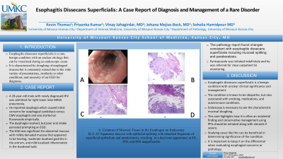
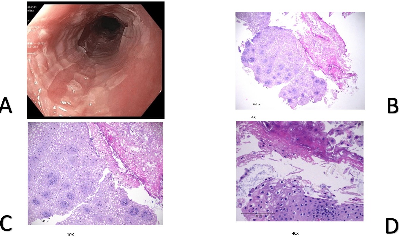
Case Description/Methods: A 28-year-old male was admitted for right lower lobe MRSA pneumonia. He was newly diagnosed with HIV. He reported dysphagia which caused initial concerns for esophageal candidiasis versus CMV esophagitis and was started on Fluconazole empirically. Though the dysphagia resolved, he continued to have poor oral intake, which prompted an EGD to be performed. The EGD was significant for abnormal mucosa with mildly denuded mucosa that appeared to be healing, moderate localized gastritis in the antrum, and mild localized inflammation in the duodenal bulb. The pathology report found changes consistent with esophagitis dissecans superficialis including mucosal splitting and parakeratosis. Pantoprazole was initiated indefinitely and he was referred for outpatient GI monitoring.
Discussion: Esophagitis dissecans superficialis is a benign condition with unclear clinical significance and management. The condition is known to be idiopathic, but also associated with smoking, medications, and autoimmune conditions. Endoscopy is necessary to see the characteristic mucosal sloughing. This case highlights how it is often an incidental finding and conservative management using PPIs should be initiated. In more severe cases, steroids should also be considered. While rare, keeping the condition on a differential diagnosis is important when evaluating a patient's esophageal concerns or pathology.

Disclosures:
Priyanka Kumar, 1, Kevin Thomas, 1, Vinay Jahagirdar, MD1, Soheila Hamidpour, MD2, Johana Meijas-Beck, MD3. P2276 - Esophagitis Dissecans Superficialis: A Digestible Introduction to a Rare Disorder, ACG 2024 Annual Scientific Meeting Abstracts. Philadelphia, PA: American College of Gastroenterology.
1University of Missouri - Kansas City School of Medicine, Kansas City, MO; 2University Health/University of Missouri, Kansas City, MO; 3University of Missouri Kansas City, Kansas City, MO
Introduction: Esophagitis dissecans superficialis is a rare, benign condition with an unclear etiology that can be visualized during an endoscopic exam. It is characterized by sloughing of esophageal mucosa but is commonly missed due to the wide variety of presentations, similarity to other conditions, and necessity of an EGD for diagnosis.
Case Description/Methods: A 28-year-old male was admitted for right lower lobe MRSA pneumonia. He was newly diagnosed with HIV. He reported dysphagia which caused initial concerns for esophageal candidiasis versus CMV esophagitis and was started on Fluconazole empirically. Though the dysphagia resolved, he continued to have poor oral intake, which prompted an EGD to be performed. The EGD was significant for abnormal mucosa with mildly denuded mucosa that appeared to be healing, moderate localized gastritis in the antrum, and mild localized inflammation in the duodenal bulb. The pathology report found changes consistent with esophagitis dissecans superficialis including mucosal splitting and parakeratosis. Pantoprazole was initiated indefinitely and he was referred for outpatient GI monitoring.
Discussion: Esophagitis dissecans superficialis is a benign condition with unclear clinical significance and management. The condition is known to be idiopathic, but also associated with smoking, medications, and autoimmune conditions. Endoscopy is necessary to see the characteristic mucosal sloughing. This case highlights how it is often an incidental finding and conservative management using PPIs should be initiated. In more severe cases, steroids should also be considered. While rare, keeping the condition on a differential diagnosis is important when evaluating a patient's esophageal concerns or pathology.

Figure: A: Columns of Mucosal Tissue in the Esophagus on Endoscopy
B, C, D: Squamous mucosa with epithelial splitting with detached fragments of superficial epithelium and parakeratosis, resulting in a dual tone appearance at 4X, 10X, and 40X magnification
B, C, D: Squamous mucosa with epithelial splitting with detached fragments of superficial epithelium and parakeratosis, resulting in a dual tone appearance at 4X, 10X, and 40X magnification
Disclosures:
Priyanka Kumar indicated no relevant financial relationships.
Kevin Thomas indicated no relevant financial relationships.
Vinay Jahagirdar indicated no relevant financial relationships.
Soheila Hamidpour indicated no relevant financial relationships.
Johana Meijas-Beck indicated no relevant financial relationships.
Priyanka Kumar, 1, Kevin Thomas, 1, Vinay Jahagirdar, MD1, Soheila Hamidpour, MD2, Johana Meijas-Beck, MD3. P2276 - Esophagitis Dissecans Superficialis: A Digestible Introduction to a Rare Disorder, ACG 2024 Annual Scientific Meeting Abstracts. Philadelphia, PA: American College of Gastroenterology.

