Monday Poster Session
Category: Esophagus
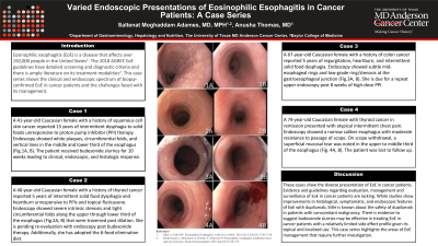
P2288 - Varied Endoscopic Presentations of Eosinophilic Esophagitis in Cancer Patients: A Case Series
Monday, October 28, 2024
10:30 AM - 4:00 PM ET
Location: Exhibit Hall E

Has Audio

Saltenat Moghaddam Adames, MD, MPH
The University of Texas MD Anderson Cancer Center, TX
Presenting Author(s)
Saltenat Moghaddam Adames, MD, MPH, Anusha Thomas, MD
The University of Texas MD Anderson Cancer Center, Houston, TX
Introduction: Eosinophilic esophagitis (EoE) is a disease that affects over 150,000 people in the United States. The 2018 AGREE EoE guidelines have detailed screening and diagnostic criteria and there is ample literature on its treatment modalities. This case series shows the endoscopic spectrum of biopsy-confirmed EoE in cancer patients and the challenges faced with its management.
Case Description/Methods:
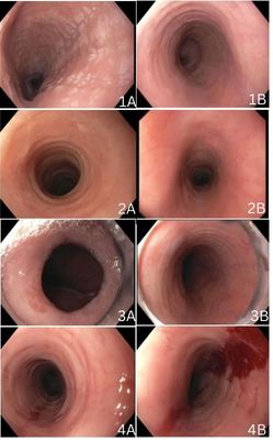
Case 1: A 41-year-old Caucasian female with a history of squamous cell skin cancer reported 15 years of intermittent dysphagia to solid foods unresponsive to proton pump inhibitor (PPI) therapy. Endoscopy showed white plaques, circumferential folds, and vertical lines in the middle and lower third of the esophagus (Fig.1A, B). The patient received budesonide slurries for 10 weeks leading to clinical, endoscopic, and histologic response.
Case 2: A 40-year-old Caucasian female with a history of thyroid cancer reported 5 years of intermittent solid food dysphagia and heartburn unresponsive to PPIs and topical fluticasone. Endoscopy showed severe intrinsic stenosis and tight circumferential folds along the upper through lower third of the esophagus (Fig.2A, B) that were traversed post dilation. She is pending re-evaluation with endoscopy post budesonide therapy. Additionally, she has adopted the 6-food elimination diet
Case 3: A 67-year-old Caucasian female with a history of colon cancer reported 5 years of regurgitation, heartburn, and intermittent solid food dysphagia. Endoscopy showed subtle mid-esophageal rings and low-grade ring/stenosis at the gastroesophageal junction (Fig.3A, B). She is due for a repeat upper endoscopy post 8 weeks of high-dose PPI.
Case 4: A 74-year-old Caucasian female with thyroid cancer in remission presented with atypical intermittent chest pain. Endoscopy showed a narrow caliber esophagus with moderate resistance to passage of scope. On scope withdrawal, a superficial mucosal tear was noted in the upper to middle third of the esophagus (Fig. 4A, B). The patient was lost to follow up.
Discussion: These cases show the diverse presentation of EoE in cancer patients. Evidence and guidelines regarding evaluation, management and surveillance of EOE in cancer patients are lacking. While studies show improvements in histological, symptomatic, and endoscopic features of EoE with dupilumab, little is known about the safety of dupilumab in patients with concomitant malignancy. This case series highlights the areas of EoE management that require further investigation.

Disclosures:
Saltenat Moghaddam Adames, MD, MPH, Anusha Thomas, MD. P2288 - Varied Endoscopic Presentations of Eosinophilic Esophagitis in Cancer Patients: A Case Series, ACG 2024 Annual Scientific Meeting Abstracts. Philadelphia, PA: American College of Gastroenterology.
The University of Texas MD Anderson Cancer Center, Houston, TX
Introduction: Eosinophilic esophagitis (EoE) is a disease that affects over 150,000 people in the United States. The 2018 AGREE EoE guidelines have detailed screening and diagnostic criteria and there is ample literature on its treatment modalities. This case series shows the endoscopic spectrum of biopsy-confirmed EoE in cancer patients and the challenges faced with its management.
Case Description/Methods:
Case 1: A 41-year-old Caucasian female with a history of squamous cell skin cancer reported 15 years of intermittent dysphagia to solid foods unresponsive to proton pump inhibitor (PPI) therapy. Endoscopy showed white plaques, circumferential folds, and vertical lines in the middle and lower third of the esophagus (Fig.1A, B). The patient received budesonide slurries for 10 weeks leading to clinical, endoscopic, and histologic response.
Case 2: A 40-year-old Caucasian female with a history of thyroid cancer reported 5 years of intermittent solid food dysphagia and heartburn unresponsive to PPIs and topical fluticasone. Endoscopy showed severe intrinsic stenosis and tight circumferential folds along the upper through lower third of the esophagus (Fig.2A, B) that were traversed post dilation. She is pending re-evaluation with endoscopy post budesonide therapy. Additionally, she has adopted the 6-food elimination diet
Case 3: A 67-year-old Caucasian female with a history of colon cancer reported 5 years of regurgitation, heartburn, and intermittent solid food dysphagia. Endoscopy showed subtle mid-esophageal rings and low-grade ring/stenosis at the gastroesophageal junction (Fig.3A, B). She is due for a repeat upper endoscopy post 8 weeks of high-dose PPI.
Case 4: A 74-year-old Caucasian female with thyroid cancer in remission presented with atypical intermittent chest pain. Endoscopy showed a narrow caliber esophagus with moderate resistance to passage of scope. On scope withdrawal, a superficial mucosal tear was noted in the upper to middle third of the esophagus (Fig. 4A, B). The patient was lost to follow up.
Discussion: These cases show the diverse presentation of EoE in cancer patients. Evidence and guidelines regarding evaluation, management and surveillance of EOE in cancer patients are lacking. While studies show improvements in histological, symptomatic, and endoscopic features of EoE with dupilumab, little is known about the safety of dupilumab in patients with concomitant malignancy. This case series highlights the areas of EoE management that require further investigation.

Figure: Figure 1A. Middle third of esophagus showing white plaques, circumferential folds, and vertical lines.
Figure 1B. Middle third of esophagus showing resolution of white plaques, circumferential folds, and vertical lines with 10 weeks of topical budesonide.
Figure 2A. Middle third of esophagus showing severe intrinsic stenosis/stricturing and tight circumferential folds.
Figure 2B. Middle third of esophagus.
Figure 3A. Low- grade rings at the gastroesophageal junction.
Figure 3B. Subtle mid-esophageal rings.
Figure 4A. Narrow caliber esophagus.
Figure 4B. Superficial mucosal tear in the upper to middle third of the esophagus.
Disclosures:
Saltenat Moghaddam Adames indicated no relevant financial relationships.
Anusha Thomas indicated no relevant financial relationships.
Saltenat Moghaddam Adames, MD, MPH, Anusha Thomas, MD. P2288 - Varied Endoscopic Presentations of Eosinophilic Esophagitis in Cancer Patients: A Case Series, ACG 2024 Annual Scientific Meeting Abstracts. Philadelphia, PA: American College of Gastroenterology.
