Monday Poster Session
Category: Esophagus
P2294 - Esophageal Granular Cell Tumor Associated With Eosinophilic Gastrointestinal Disease: A Case Report
Monday, October 28, 2024
10:30 AM - 4:00 PM ET
Location: Exhibit Hall E

Has Audio

Cathy Zheng, MD
University of Miami Miller School of Medicine at Jackson Memorial Hospital
Miami, FL
Presenting Author(s)
Cathy Zheng, MD1, Robert Mowery, DO2, Sareena Ali, DO3, Ryan T. Hoff, DO4
1University of Miami Miller School of Medicine at Jackson Memorial Hospital, Miami, FL; 2Advocate Lutheran General Hospital, Chicago, IL; 3Advocate Lutheran General Hospital, Park Ridge, IL; 4PeaceHealth Southwest Medical Center, Vancouver, WA
Introduction: There is a possible link between eosinophilic esophagitis (EoE) and granular cell tumors (GCT). We present a rare case of esophageal GCT diagnosed in a patient with a long history of eosinophilic gastrointestinal disease (EGID) and likely several years of active EoE preceding the GCT.
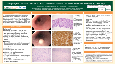
Case Description/Methods: A 33-year-old woman with a reported distant history of EGID diagnosed by esophagogastroduodenoscopy (EGD) presented with chronic regurgitation and progressive intermittent dysphagia to solid foods. She was treated with a short course of intravenous steroids upon diagnosis of EGID about 12 years prior but had otherwise remained off therapy. EGD showed esophageal edema, longitudinal furrows and exudates, a sessile nodule in the mid esophagus, and duodenal erythema. Biopsies showed eosinophilic gastroduodenitis with esophageal involvement. Biopsy of the mid esophageal lesion revealed a granular cell tumor.
The patient started a six-food elimination diet and reported symptom improvement after 8 weeks. Follow-up EGD at 8 weeks re-demonstrated a nodule in the mid esophagus, which was removed en bloc via endoscopic mucosal resection. EGD biopsies showed resolution of previously prominent eosinophilic gastroduodenitis and improved but persistent EoE (5 eosinophils per high-power field (hpf) in the distal esophagus and 20 per hpf in the mid esophagus). The patient proceeded with reintroduction as a part of the six-food elimination diet. At the second follow-up EGD 4 weeks later, she continued to report good control of symptoms. EGD and biopsies were consistent with remission (12 eosinophils per hpf in the proximal and distal esophagus).
Discussion: GCTs are typically benign lesions thought to be of Schwann cell origin. About 1% of GCTs are located in the esophagus. GCTs are often asymptomatic, though larger lesions can cause dysphagia, chest pain, cough, nausea, and gastroesophageal reflux. Malignant transformation occurs in 1-2% of cases.
We present a rare case of esophageal GCT in a patient with a remote history of EGID. EoE is a chronic, immune/antigen-mediated disease characterized histologically by eosinophil-predominant inflammation that causes symptoms of esophageal dysfunction. One hypothesis is that eosinophilic inflammation may increase the risk of esophageal GCT formation, as GCTs have previously been linked to sites of scarring and inflammation. Our case suggests an association between esophageal GCT and EoE, but the underlying pathogenesis warrants further investigation.

Disclosures:
Cathy Zheng, MD1, Robert Mowery, DO2, Sareena Ali, DO3, Ryan T. Hoff, DO4. P2294 - Esophageal Granular Cell Tumor Associated With Eosinophilic Gastrointestinal Disease: A Case Report, ACG 2024 Annual Scientific Meeting Abstracts. Philadelphia, PA: American College of Gastroenterology.
1University of Miami Miller School of Medicine at Jackson Memorial Hospital, Miami, FL; 2Advocate Lutheran General Hospital, Chicago, IL; 3Advocate Lutheran General Hospital, Park Ridge, IL; 4PeaceHealth Southwest Medical Center, Vancouver, WA
Introduction: There is a possible link between eosinophilic esophagitis (EoE) and granular cell tumors (GCT). We present a rare case of esophageal GCT diagnosed in a patient with a long history of eosinophilic gastrointestinal disease (EGID) and likely several years of active EoE preceding the GCT.
Case Description/Methods: A 33-year-old woman with a reported distant history of EGID diagnosed by esophagogastroduodenoscopy (EGD) presented with chronic regurgitation and progressive intermittent dysphagia to solid foods. She was treated with a short course of intravenous steroids upon diagnosis of EGID about 12 years prior but had otherwise remained off therapy. EGD showed esophageal edema, longitudinal furrows and exudates, a sessile nodule in the mid esophagus, and duodenal erythema. Biopsies showed eosinophilic gastroduodenitis with esophageal involvement. Biopsy of the mid esophageal lesion revealed a granular cell tumor.
The patient started a six-food elimination diet and reported symptom improvement after 8 weeks. Follow-up EGD at 8 weeks re-demonstrated a nodule in the mid esophagus, which was removed en bloc via endoscopic mucosal resection. EGD biopsies showed resolution of previously prominent eosinophilic gastroduodenitis and improved but persistent EoE (5 eosinophils per high-power field (hpf) in the distal esophagus and 20 per hpf in the mid esophagus). The patient proceeded with reintroduction as a part of the six-food elimination diet. At the second follow-up EGD 4 weeks later, she continued to report good control of symptoms. EGD and biopsies were consistent with remission (12 eosinophils per hpf in the proximal and distal esophagus).
Discussion: GCTs are typically benign lesions thought to be of Schwann cell origin. About 1% of GCTs are located in the esophagus. GCTs are often asymptomatic, though larger lesions can cause dysphagia, chest pain, cough, nausea, and gastroesophageal reflux. Malignant transformation occurs in 1-2% of cases.
We present a rare case of esophageal GCT in a patient with a remote history of EGID. EoE is a chronic, immune/antigen-mediated disease characterized histologically by eosinophil-predominant inflammation that causes symptoms of esophageal dysfunction. One hypothesis is that eosinophilic inflammation may increase the risk of esophageal GCT formation, as GCTs have previously been linked to sites of scarring and inflammation. Our case suggests an association between esophageal GCT and EoE, but the underlying pathogenesis warrants further investigation.

Figure: A. Image from initial upper endoscopy showing esophageal granular cell tumor at 31 cm from the incisors, which appears sessile and white. Also present are esophageal exudates.
B. Histologic image of the granular cell tumor showing overlying squamous mucosa. Epithelioid granular cell tumors are characterized by their rounded appearance and large cytoplasm containing eosinophilic granules and numerous lysosomes. Note that the tumor does not invade the mucosa; no aggressive features were present.
C. Image showing S-100 immunohistochemical stain with 100x magnification, confirming the diagnosis of GCT.
Disclosures:
Cathy Zheng indicated no relevant financial relationships.
Robert Mowery indicated no relevant financial relationships.
Sareena Ali indicated no relevant financial relationships.
Ryan Hoff indicated no relevant financial relationships.
Cathy Zheng, MD1, Robert Mowery, DO2, Sareena Ali, DO3, Ryan T. Hoff, DO4. P2294 - Esophageal Granular Cell Tumor Associated With Eosinophilic Gastrointestinal Disease: A Case Report, ACG 2024 Annual Scientific Meeting Abstracts. Philadelphia, PA: American College of Gastroenterology.
