Monday Poster Session
Category: Esophagus
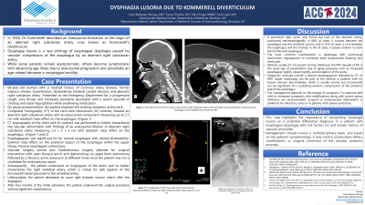
P2301 - Dysphagia Lusoria Due to Kommerell Diverticulum
Monday, October 28, 2024
10:30 AM - 4:00 PM ET
Location: Exhibit Hall E

Has Audio

Carla Barberan Parraga, MD
Maimonides Medical Center
New York, NY
Presenting Author(s)
Carla Barberan Parraga, MD1, Tanuj Chokshi, DO2, Palvi Singla, MBBS, MD3, Seth Lapin, DO3
1Maimonides Medical Center, New York, NY; 2Maimonides Medical Center, Clifton, NJ; 3Maimonides Medical Center, Brooklyn, NY
Introduction: In 1936, Dr. Kommerell described an aneurysmal formation at the origin of an aberrant right subclavian artery, now known as Kommerell’s diverticulum. Dysphagia lusoria is a rare etiology of esophageal dysphagia caused by vascular compression of the esophagus by an aberrant right subclavian artery. This is the most common vascular congenital abnormality of the aortic arch. While some patients remain asymptomatic, others become symptomatic with advancing age, likely due to aneurysmal progression and potentially an age-related decrease in esophageal motility.
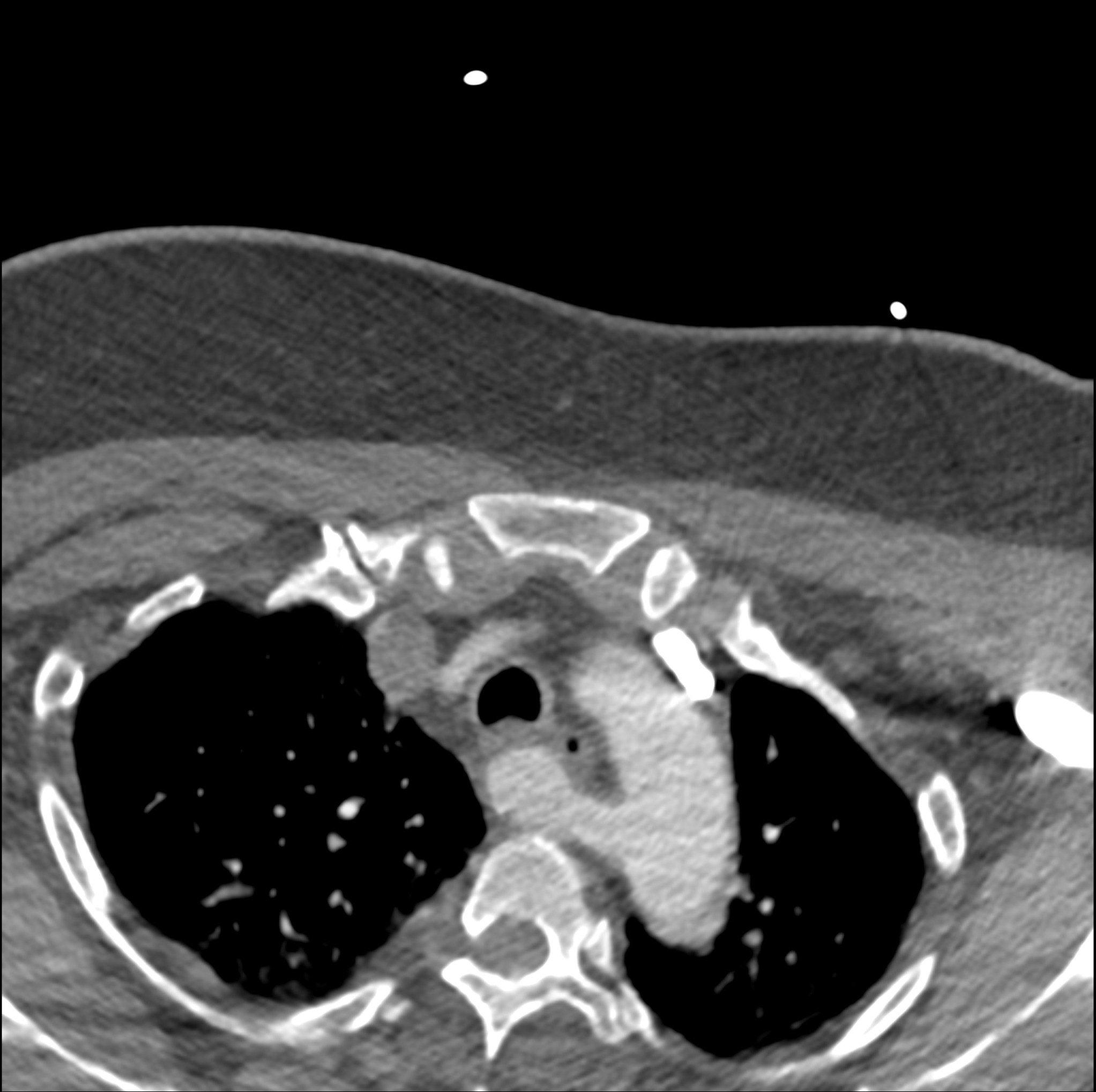
Case Description/Methods: Here, we describe a case of a 64-year-old woman with a medical history of obesity, coronary artery disease, former tobacco smoking, hypertension, dyslipidemia, bilateral carotid stenosis, and aberrant right subclavian artery. The patient presented to the Emergency Department for a progressive cough, sore throat, and increased secretions associated with a recent episode of choking and nasal regurgitation in an attempt to swallow medication. On physical examination, the patient localized the choking sensation at the neck. Further workup, including a Computed Tomography (CT) of the neck with intravenous (IV) contrast, showed an aberrant right subclavian artery with an aneurysmal component measuring up to 2.1 cm with a resultant mass effect on the esophagus. A CT angiogram of the chest with IV contrast was performed to further characterize the vascular abnormality with findings of an aneurysmal dilation of the aberrant right subclavian artery measuring 2.5 x 2 x 2 cm with a resultant mass effect on the esophagus. Vascular Surgery planned for surgical intervention with open thoracic aortic vessel debranching since the patient was not a candidate for endovascular repair.
Discussion: This case highlights the importance of recognizing dysphagia lusoria as a potential differential diagnosis in a patient with esophageal dysphagia with risk factors for and known history of vascular anomalies. Diagnostic workup should include a barium esophagogram and computed tomography or magnetic resonance angiography. Management should involve a multidisciplinary team and can range from conservative dietary modifications to surgical correction of the vascular anatomic anomaly based on the patient’s symptomatology and comorbidities.

Disclosures:
Carla Barberan Parraga, MD1, Tanuj Chokshi, DO2, Palvi Singla, MBBS, MD3, Seth Lapin, DO3. P2301 - Dysphagia Lusoria Due to Kommerell Diverticulum, ACG 2024 Annual Scientific Meeting Abstracts. Philadelphia, PA: American College of Gastroenterology.
1Maimonides Medical Center, New York, NY; 2Maimonides Medical Center, Clifton, NJ; 3Maimonides Medical Center, Brooklyn, NY
Introduction: In 1936, Dr. Kommerell described an aneurysmal formation at the origin of an aberrant right subclavian artery, now known as Kommerell’s diverticulum. Dysphagia lusoria is a rare etiology of esophageal dysphagia caused by vascular compression of the esophagus by an aberrant right subclavian artery. This is the most common vascular congenital abnormality of the aortic arch. While some patients remain asymptomatic, others become symptomatic with advancing age, likely due to aneurysmal progression and potentially an age-related decrease in esophageal motility.
Case Description/Methods: Here, we describe a case of a 64-year-old woman with a medical history of obesity, coronary artery disease, former tobacco smoking, hypertension, dyslipidemia, bilateral carotid stenosis, and aberrant right subclavian artery. The patient presented to the Emergency Department for a progressive cough, sore throat, and increased secretions associated with a recent episode of choking and nasal regurgitation in an attempt to swallow medication. On physical examination, the patient localized the choking sensation at the neck. Further workup, including a Computed Tomography (CT) of the neck with intravenous (IV) contrast, showed an aberrant right subclavian artery with an aneurysmal component measuring up to 2.1 cm with a resultant mass effect on the esophagus. A CT angiogram of the chest with IV contrast was performed to further characterize the vascular abnormality with findings of an aneurysmal dilation of the aberrant right subclavian artery measuring 2.5 x 2 x 2 cm with a resultant mass effect on the esophagus. Vascular Surgery planned for surgical intervention with open thoracic aortic vessel debranching since the patient was not a candidate for endovascular repair.
Discussion: This case highlights the importance of recognizing dysphagia lusoria as a potential differential diagnosis in a patient with esophageal dysphagia with risk factors for and known history of vascular anomalies. Diagnostic workup should include a barium esophagogram and computed tomography or magnetic resonance angiography. Management should involve a multidisciplinary team and can range from conservative dietary modifications to surgical correction of the vascular anatomic anomaly based on the patient’s symptomatology and comorbidities.

Figure: CTA head and neck with IV contrast, with aberrant right subclavian artery with associated aneurysmal dilation compressing the esophagus.
Disclosures:
Carla Barberan Parraga indicated no relevant financial relationships.
Tanuj Chokshi indicated no relevant financial relationships.
Palvi Singla indicated no relevant financial relationships.
Seth Lapin indicated no relevant financial relationships.
Carla Barberan Parraga, MD1, Tanuj Chokshi, DO2, Palvi Singla, MBBS, MD3, Seth Lapin, DO3. P2301 - Dysphagia Lusoria Due to Kommerell Diverticulum, ACG 2024 Annual Scientific Meeting Abstracts. Philadelphia, PA: American College of Gastroenterology.
