Monday Poster Session
Category: General Endoscopy
P2412 - Stop Pushing Too Hard! Challenging Colonoscopy May Indicate Unexpected Pathology
Monday, October 28, 2024
10:30 AM - 4:00 PM ET
Location: Exhibit Hall E

Has Audio

Keming Yang, MD, PhD
University of Pittsburgh Medical Center
Pittsburgh, PA
Presenting Author(s)
Keming Yang, MD, PhD, Robert Schoen, MD, MS
University of Pittsburgh Medical Center, Pittsburgh, PA
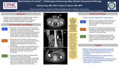
Introduction: Complete colonoscopy is essential for cancer screening. However, complete colonoscopy is not always possible. We share three incomplete colonoscopies that were challenged by unexpected pathology revealed during subsequent CT colonography.
Case Description/Methods: Case A: A 83-year-old man with a family history of colon cancer presented for a screening colonoscopy. The patient had multiple prior successful colonoscopy exams. The current colonoscopy was unusually difficult due to a tortuous colon with a sharp angulation in the left colon. Despite multiple maneuvers and position change, the scope could not successfully navigate around that turn.
Case B: A 55-year-old man with a history of rectal cancer s/p resection and colo-colonic anastomosis presented for a surveillance colonoscopy. A previous exam was described as extremely difficult and required a slim colonoscope. This colonoscopy was also extremely difficult due to a tortuous colon with a sharp, angulated turn near the splenic flexure. Attempts at completion via position change and use of a slim colonoscope were unsuccessful.
Case C: A 65-year-old woman with a history of cholecystectomy presented for a screening colonoscopy. The colonoscopy was technically difficult and complex due to unusual anatomy with a sharp turn at 30cm that could not be visualized/crossed despite rotating her position and changing to a more flexible scope. The scope was only advanced to the sigmoid colon.
Discussion: Decision and Findings: All three patients underwent CT colonography.
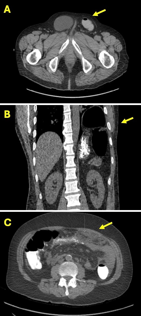
Case A: Same day CT colonography showed a left inguinal hernia with sigmoid colon herniation within the hernia sac (Fig1A). This required subsequent surgical repair.
Case B: Same day CT colonography showed herniation of the splenic flexure through a left sided Bochdalek diaphragmatic defect (Fig1B). The defect required subsequent thoracoscopic repair.
Case C: Subsequent CT colonography showed a likely ovarian cancer with peritoneal and sigmoid colon extrinsic involvement (Fig1C). Patient is currently under evaluation by the gynecologic oncology service.
Lesson: Difficult colonoscopy may indicate unexpected findings. Excessive pushing during such cases could incur severe adverse events such as perforation. CT colonography should be considered when incomplete colonoscopy occurs. Had excessive insertion force been applied in these cases, it would almost certainly have resulted in catastrophic complications.

Disclosures:
Keming Yang, MD, PhD, Robert Schoen, MD, MS. P2412 - Stop Pushing Too Hard! Challenging Colonoscopy May Indicate Unexpected Pathology, ACG 2024 Annual Scientific Meeting Abstracts. Philadelphia, PA: American College of Gastroenterology.
University of Pittsburgh Medical Center, Pittsburgh, PA
Introduction: Complete colonoscopy is essential for cancer screening. However, complete colonoscopy is not always possible. We share three incomplete colonoscopies that were challenged by unexpected pathology revealed during subsequent CT colonography.
Case Description/Methods: Case A: A 83-year-old man with a family history of colon cancer presented for a screening colonoscopy. The patient had multiple prior successful colonoscopy exams. The current colonoscopy was unusually difficult due to a tortuous colon with a sharp angulation in the left colon. Despite multiple maneuvers and position change, the scope could not successfully navigate around that turn.
Case B: A 55-year-old man with a history of rectal cancer s/p resection and colo-colonic anastomosis presented for a surveillance colonoscopy. A previous exam was described as extremely difficult and required a slim colonoscope. This colonoscopy was also extremely difficult due to a tortuous colon with a sharp, angulated turn near the splenic flexure. Attempts at completion via position change and use of a slim colonoscope were unsuccessful.
Case C: A 65-year-old woman with a history of cholecystectomy presented for a screening colonoscopy. The colonoscopy was technically difficult and complex due to unusual anatomy with a sharp turn at 30cm that could not be visualized/crossed despite rotating her position and changing to a more flexible scope. The scope was only advanced to the sigmoid colon.
Discussion: Decision and Findings: All three patients underwent CT colonography.
Case A: Same day CT colonography showed a left inguinal hernia with sigmoid colon herniation within the hernia sac (Fig1A). This required subsequent surgical repair.
Case B: Same day CT colonography showed herniation of the splenic flexure through a left sided Bochdalek diaphragmatic defect (Fig1B). The defect required subsequent thoracoscopic repair.
Case C: Subsequent CT colonography showed a likely ovarian cancer with peritoneal and sigmoid colon extrinsic involvement (Fig1C). Patient is currently under evaluation by the gynecologic oncology service.
Lesson: Difficult colonoscopy may indicate unexpected findings. Excessive pushing during such cases could incur severe adverse events such as perforation. CT colonography should be considered when incomplete colonoscopy occurs. Had excessive insertion force been applied in these cases, it would almost certainly have resulted in catastrophic complications.

Figure: Fig 1. Unexpected pathology revealed during subsequent CT colonography following incomplete colonoscopy.
A: left inguinal hernia with sigmoid colon herniated within the hernia. B: herniation of the splenic flexure through a left sided Bochdalek diaphragmatic defect. C: peritoneal and sigmoid colon extrinsic involvement from ovarian cancer.
A: left inguinal hernia with sigmoid colon herniated within the hernia. B: herniation of the splenic flexure through a left sided Bochdalek diaphragmatic defect. C: peritoneal and sigmoid colon extrinsic involvement from ovarian cancer.
Disclosures:
Keming Yang indicated no relevant financial relationships.
Robert Schoen: Exact Sciences – Grant/Research Support. Freenome – Grant/Research Support. Guardant – Advisory Committee/Board Member. Immunovia. Inc – Grant/Research Support.
Keming Yang, MD, PhD, Robert Schoen, MD, MS. P2412 - Stop Pushing Too Hard! Challenging Colonoscopy May Indicate Unexpected Pathology, ACG 2024 Annual Scientific Meeting Abstracts. Philadelphia, PA: American College of Gastroenterology.
