Monday Poster Session
Category: General Endoscopy
P2420 - A Rare Case of Appendiceal Intussusception Secondary to Endometriosis
Monday, October 28, 2024
10:30 AM - 4:00 PM ET
Location: Exhibit Hall E

Has Audio

Jayashrei Sairam, MD
Greater Baltimore Medical Center
Towson, MD
Presenting Author(s)
Jayashrei Sairam, MD1, Akshay Duddu, MD1, Nadim Jaafar, MD1, M Kenan Rahima, MD2, Beatriz Briones, MD1
1Greater Baltimore Medical Center, Towson, MD; 2TriHealth Good Samaritan Hospital, Cincinnati, OH
Introduction: Appendiceal intussusception (AI) is a rare condition with an incidence of 0.01%. Endometriosis is an even rare cause of AI, with literature limited to a few case reports. AI constitutes a diagnostic challenge for physicians, with the diagnosis primarily made intraoperatively or postoperatively. We report a case of AI secondary to endometriosis in a patient with no history of endometriosis.
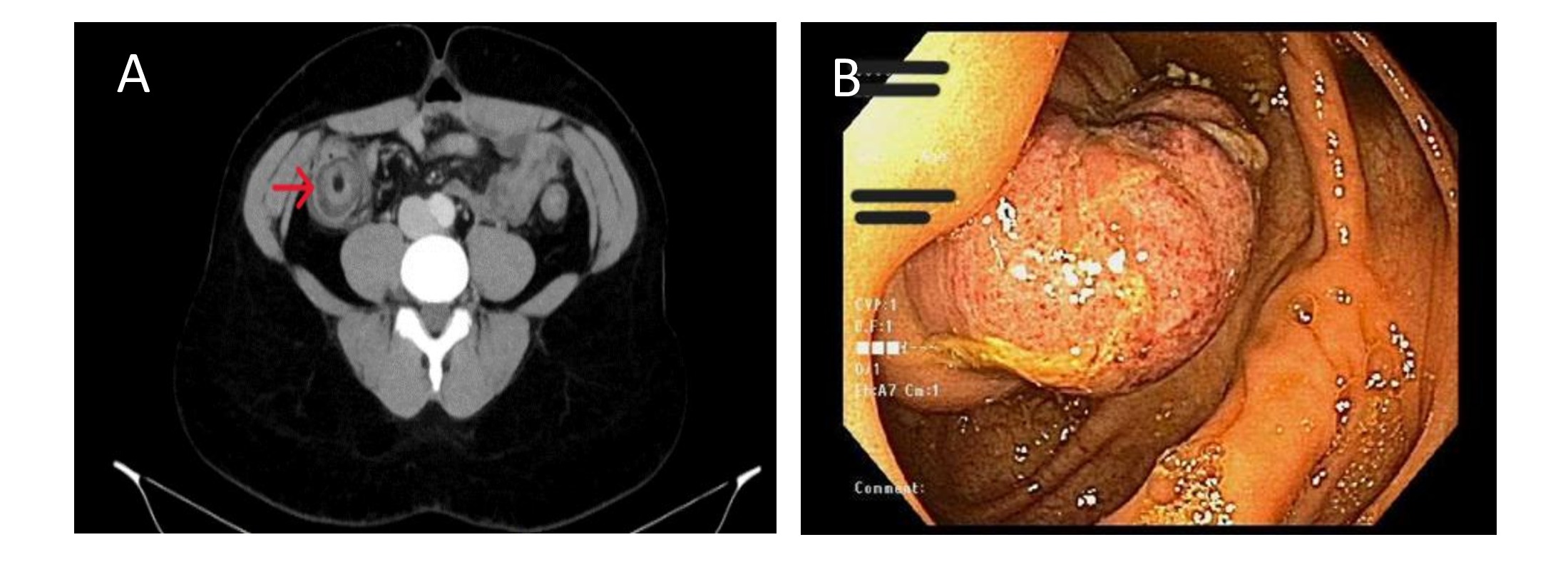
Case Description/Methods: A 38-year-old lady with no past medical history presented with a 2-day history of diffuse abdominal pain and vomiting. A CT revealed a “target” lesion at the ileocecal region with the appearance of the terminal ileum telescoping into the ascending colon, concerning for an ileocolic intussusception. A fluoroscopic small bowel follow-through series showed no signs of small bowel obstruction. A colonoscopy performed in an attempt to reduce the intussusception revealed a polypoid non-obstructing lesion at the appendiceal orifice, suspicious for an appendiceal mass, or an inverted appendix. Histopathology returned negative for malignancy. Exploratory laparoscopy showed AI with an inverted appendix invaginating into the caecum, unable to be reduced. The patient underwent laparoscopic wedge cecectomy and appendectomy. Histopathology revealed an inverted appendix with ischemic changes, fibrosis and serosal adhesions of the appendiceal tip secondary to endometriosis.
Discussion: AI is a condition characterized by appendiceal hyperperistalsis secondary to anatomical or pathological processes leading to invagination into the caecum. The incidence of gastrointestinal endometriosis varies between 3%-37%, with the rectum and sigmoid colon being the most common sites. Appendiceal involvement is seen in only 3% of the cases. The chronic serosal fibrosis with eventual hypertrophy of the muscularis propria seen in endometriosis acts as a pathological lead point, making it prone to intussusception. Preoperative diagnosis is rare; however, if imaging suggests the diagnosis, a colonoscopy allows for direct visualization of the lesion. The findings may, however mimic a cecal mass and are often mistaken for a neoplasm. Definitive treatment is with a partial cecectomy and appendectomy. Recognition of AI by endoscopists is crucial to avoid misdiagnosis, as this is commonly mistaken for a polyp/mass. Misguided attempts at endoscopic removal of the lesion or inappropriate surgery can potentially lead to complications like perforation or peritonitis.

Disclosures:
Jayashrei Sairam, MD1, Akshay Duddu, MD1, Nadim Jaafar, MD1, M Kenan Rahima, MD2, Beatriz Briones, MD1. P2420 - A Rare Case of Appendiceal Intussusception Secondary to Endometriosis, ACG 2024 Annual Scientific Meeting Abstracts. Philadelphia, PA: American College of Gastroenterology.
1Greater Baltimore Medical Center, Towson, MD; 2TriHealth Good Samaritan Hospital, Cincinnati, OH
Introduction: Appendiceal intussusception (AI) is a rare condition with an incidence of 0.01%. Endometriosis is an even rare cause of AI, with literature limited to a few case reports. AI constitutes a diagnostic challenge for physicians, with the diagnosis primarily made intraoperatively or postoperatively. We report a case of AI secondary to endometriosis in a patient with no history of endometriosis.
Case Description/Methods: A 38-year-old lady with no past medical history presented with a 2-day history of diffuse abdominal pain and vomiting. A CT revealed a “target” lesion at the ileocecal region with the appearance of the terminal ileum telescoping into the ascending colon, concerning for an ileocolic intussusception. A fluoroscopic small bowel follow-through series showed no signs of small bowel obstruction. A colonoscopy performed in an attempt to reduce the intussusception revealed a polypoid non-obstructing lesion at the appendiceal orifice, suspicious for an appendiceal mass, or an inverted appendix. Histopathology returned negative for malignancy. Exploratory laparoscopy showed AI with an inverted appendix invaginating into the caecum, unable to be reduced. The patient underwent laparoscopic wedge cecectomy and appendectomy. Histopathology revealed an inverted appendix with ischemic changes, fibrosis and serosal adhesions of the appendiceal tip secondary to endometriosis.
Discussion: AI is a condition characterized by appendiceal hyperperistalsis secondary to anatomical or pathological processes leading to invagination into the caecum. The incidence of gastrointestinal endometriosis varies between 3%-37%, with the rectum and sigmoid colon being the most common sites. Appendiceal involvement is seen in only 3% of the cases. The chronic serosal fibrosis with eventual hypertrophy of the muscularis propria seen in endometriosis acts as a pathological lead point, making it prone to intussusception. Preoperative diagnosis is rare; however, if imaging suggests the diagnosis, a colonoscopy allows for direct visualization of the lesion. The findings may, however mimic a cecal mass and are often mistaken for a neoplasm. Definitive treatment is with a partial cecectomy and appendectomy. Recognition of AI by endoscopists is crucial to avoid misdiagnosis, as this is commonly mistaken for a polyp/mass. Misguided attempts at endoscopic removal of the lesion or inappropriate surgery can potentially lead to complications like perforation or peritonitis.

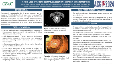
Figure: Figure A: Axial view of CT showing target sign, concerning for ileocolic intussusception.
Figure B: Colonoscopy showing a polypoid non-obstructing medium-sized mass invaginating into the cecum.
Figure B: Colonoscopy showing a polypoid non-obstructing medium-sized mass invaginating into the cecum.
Disclosures:
Jayashrei Sairam indicated no relevant financial relationships.
Akshay Duddu indicated no relevant financial relationships.
Nadim Jaafar indicated no relevant financial relationships.
M Kenan Rahima indicated no relevant financial relationships.
Beatriz Briones indicated no relevant financial relationships.
Jayashrei Sairam, MD1, Akshay Duddu, MD1, Nadim Jaafar, MD1, M Kenan Rahima, MD2, Beatriz Briones, MD1. P2420 - A Rare Case of Appendiceal Intussusception Secondary to Endometriosis, ACG 2024 Annual Scientific Meeting Abstracts. Philadelphia, PA: American College of Gastroenterology.

