Monday Poster Session
Category: General Endoscopy
P2434 - Glass Half Empty or Half Full: Single Session Endoscopic Removal of Multiple Glass Vials in the Upper Gastrointestinal Tract Using a Novel Approach
Monday, October 28, 2024
10:30 AM - 4:00 PM ET
Location: Exhibit Hall E

Has Audio

Raghav Bassi, MD
University of Central Florida College of Medicine
Gainesville, FL
Presenting Author(s)
Raghav Bassi, MD1, Gabriel S.. Silva, MD2, Shreyans Doshi, MD3, Tony S.. Brar, MD1, Yaseen Perbtani, DO1
1University of Central Florida College of Medicine, Gainesville, FL; 2Aventura Hospital, Aventura, FL; 3Wake Forest University School of Medicine, Charlotte, NC
Introduction: In adults, foreign body impaction is frequently seen in the setting of bone or meat bolus impaction from underlying structural pathologies such as esophageal rings, achalasia, webs, benign or malignant strictures, or eosinophilic esophagitis. 80-90% of ingested foreign bodies will pass spontaneously, with only 10-20% necessitating endoscopic removal and less than 1% needing surgical intervention.
Case Description/Methods: A 23-year-old transgender female presented to the emergency department with epigastric tenderness and non-bloody but bilious emesis that started one day before admission. On presentation, there was no evidence of guarding or rebound tenderness. An abdominal x-ray (KUB) was done and it revealed close to thirty radiopaque densities in the stomach, with some extending to the duodenum and distal colon (Figure 1). An emergent esophagogastroduodenoscopy was planned for the endoscopic removal of these objects. During the procedure, an overtube was placed to help facilitate their removal, however, the inner diameter was too small to accommodate the transoral removal of the vials. Multiple retrieval tools were utilized including a polypectomy snare, retrieval basket, and alligator forceps, however, given the smooth round edges, the vials were not able to transverse through the upper esophageal sphincter. A novel approach was utilized through a combination of both a snare and a 15mm extraction balloon. The 15mm extraction balloon was inflated inside the lumen of the glass vials to increase the intraluminal pressure and allow for the safe removal of these vials individually. Twenty-seven 6cm x1cm glass vials were successfully and safely removed from the stomach and proximal duodenum. A repeat KUB post-procedure revealed three glass vials that migrated to the ascending colon with plans for a colonoscopy the next day if the vials failed to pass spontaneously. The patient felt better with a complete resolution of her symptoms.
Discussion: Long slender objects greater than 5 cm long, have a difficult time traversing through the gastrointestional tract and have an increased likelihood of getting lodged in the duodenal sweep. Fortunately, endoscopists can deploy a wide array of retrieval tools depending on the features of the foreign object. As seen in our case above, the round smooth edges of the glass vial made it difficult to grasp the glass vials using traditional techniques, however, a combination of snare and a 15mm extraction balloon was successful in the removal of these vials.

Disclosures:
Raghav Bassi, MD1, Gabriel S.. Silva, MD2, Shreyans Doshi, MD3, Tony S.. Brar, MD1, Yaseen Perbtani, DO1. P2434 - Glass Half Empty or Half Full: Single Session Endoscopic Removal of Multiple Glass Vials in the Upper Gastrointestinal Tract Using a Novel Approach, ACG 2024 Annual Scientific Meeting Abstracts. Philadelphia, PA: American College of Gastroenterology.
1University of Central Florida College of Medicine, Gainesville, FL; 2Aventura Hospital, Aventura, FL; 3Wake Forest University School of Medicine, Charlotte, NC
Introduction: In adults, foreign body impaction is frequently seen in the setting of bone or meat bolus impaction from underlying structural pathologies such as esophageal rings, achalasia, webs, benign or malignant strictures, or eosinophilic esophagitis. 80-90% of ingested foreign bodies will pass spontaneously, with only 10-20% necessitating endoscopic removal and less than 1% needing surgical intervention.
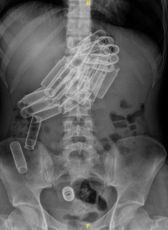
Case Description/Methods: A 23-year-old transgender female presented to the emergency department with epigastric tenderness and non-bloody but bilious emesis that started one day before admission. On presentation, there was no evidence of guarding or rebound tenderness. An abdominal x-ray (KUB) was done and it revealed close to thirty radiopaque densities in the stomach, with some extending to the duodenum and distal colon (Figure 1). An emergent esophagogastroduodenoscopy was planned for the endoscopic removal of these objects. During the procedure, an overtube was placed to help facilitate their removal, however, the inner diameter was too small to accommodate the transoral removal of the vials. Multiple retrieval tools were utilized including a polypectomy snare, retrieval basket, and alligator forceps, however, given the smooth round edges, the vials were not able to transverse through the upper esophageal sphincter. A novel approach was utilized through a combination of both a snare and a 15mm extraction balloon. The 15mm extraction balloon was inflated inside the lumen of the glass vials to increase the intraluminal pressure and allow for the safe removal of these vials individually. Twenty-seven 6cm x1cm glass vials were successfully and safely removed from the stomach and proximal duodenum. A repeat KUB post-procedure revealed three glass vials that migrated to the ascending colon with plans for a colonoscopy the next day if the vials failed to pass spontaneously. The patient felt better with a complete resolution of her symptoms.
Discussion: Long slender objects greater than 5 cm long, have a difficult time traversing through the gastrointestional tract and have an increased likelihood of getting lodged in the duodenal sweep. Fortunately, endoscopists can deploy a wide array of retrieval tools depending on the features of the foreign object. As seen in our case above, the round smooth edges of the glass vial made it difficult to grasp the glass vials using traditional techniques, however, a combination of snare and a 15mm extraction balloon was successful in the removal of these vials.

Figure: Fig 1: Abdominal x-ray revealing multiple glass vials in the stomach with extension to the duodenum and one vial in the distal colon.
Disclosures:
Raghav Bassi indicated no relevant financial relationships.
Gabriel Silva indicated no relevant financial relationships.
Shreyans Doshi indicated no relevant financial relationships.
Tony Brar indicated no relevant financial relationships.
Yaseen Perbtani indicated no relevant financial relationships.
Raghav Bassi, MD1, Gabriel S.. Silva, MD2, Shreyans Doshi, MD3, Tony S.. Brar, MD1, Yaseen Perbtani, DO1. P2434 - Glass Half Empty or Half Full: Single Session Endoscopic Removal of Multiple Glass Vials in the Upper Gastrointestinal Tract Using a Novel Approach, ACG 2024 Annual Scientific Meeting Abstracts. Philadelphia, PA: American College of Gastroenterology.
