Monday Poster Session
Category: General Endoscopy
P2436 - Transhepatic Percutaneous Endoscopic Gastrostomy Tube: A Rare and Underrecognized Complication
Monday, October 28, 2024
10:30 AM - 4:00 PM ET
Location: Exhibit Hall E

Has Audio

Osama Alshakhatreh, MD
Albany Medical Center
Albany, NY
Presenting Author(s)
Osama Alshakhatreh, MD, Khaled Elsokary, DO, Omar Tageldin, MD, Joseph Soucy, MD, Robert Gianotti, MD
Albany Medical Center, Albany, NY
Introduction: Percutaneous endoscopic gastrostomy (PEG) tube placement is a common procedure with multiple techniques described, including the pull, push, and introducer PEG. Regardless of the method used, PEG tube placement involves the blind perforation of the gastrointestinal system through a skin incision, thus risking injury to intrabdominal organs. Transhepatic PEG tube placement is a rare complication. It happens when the left lobe of the liver lies between the stomach and the abdominal wall, rendering it susceptible to injury.
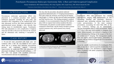
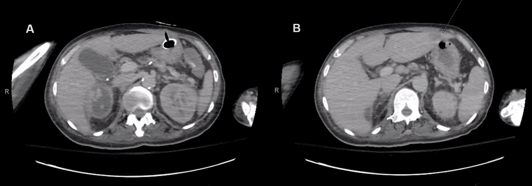
Case Description/Methods: A 56-year-old male presented with back pain and weakness after a fall. He was found to have septic shock due to a urinary tract infection, necessitating pressure and respiratory support. Following improvement, he underwent PEG tube placement for pharyngeal dysphagia. Using the pull technique, a 24F PEG tube was placed with excellent endoscopic transillumination and one-one indentation. One day after the procedure, the patient endorsed generalized abdominal pain. CT imaging demonstrated a PEG tube within the stomach, traversing the left hepatic lobe [Figure 1]. Follow-up labs showed stable hemoglobin and liver function tests. This finding prompted a surgical evaluation. It was recommended to keep the PEG tube in place with no surgical intervention needed at the time. Unfortunately, the patient's overall medical condition continued to deteriorate, and the family decided to pursue comfort measures only, and he passed away shortly afterward.
Discussion: Transhepatic PEG tube placement has variable presentations, ranging from asymptomatic to life-threatening bleeding and peritonitis. However, abdominal pain, presenting anytime from hours to weeks post-procedure, is the most common presentation, accounting for more than 50% of the cases. Laboratory findings can be unremarkable, showing hemoglobin drop or mild transaminitis. While a CT scan is the most used modality to diagnose liver injury, ultrasonography was utilized to diagnose a few cases, demonstrating findings at the margins of the liver not evident on CT imaging. The management usually does not require surgical intervention, particularly in patients with no evidence of significant liver lacerations or bleeding.
Given the variable manifestations of a transhepatic PEG tube, it may be an underrecognized complication. It is important to keep a low threshold to suspect hepatic injury in patients with subtle symptoms following PEG tube placement.

Disclosures:
Osama Alshakhatreh, MD, Khaled Elsokary, DO, Omar Tageldin, MD, Joseph Soucy, MD, Robert Gianotti, MD. P2436 - Transhepatic Percutaneous Endoscopic Gastrostomy Tube: A Rare and Underrecognized Complication, ACG 2024 Annual Scientific Meeting Abstracts. Philadelphia, PA: American College of Gastroenterology.
Albany Medical Center, Albany, NY
Introduction: Percutaneous endoscopic gastrostomy (PEG) tube placement is a common procedure with multiple techniques described, including the pull, push, and introducer PEG. Regardless of the method used, PEG tube placement involves the blind perforation of the gastrointestinal system through a skin incision, thus risking injury to intrabdominal organs. Transhepatic PEG tube placement is a rare complication. It happens when the left lobe of the liver lies between the stomach and the abdominal wall, rendering it susceptible to injury.
Case Description/Methods: A 56-year-old male presented with back pain and weakness after a fall. He was found to have septic shock due to a urinary tract infection, necessitating pressure and respiratory support. Following improvement, he underwent PEG tube placement for pharyngeal dysphagia. Using the pull technique, a 24F PEG tube was placed with excellent endoscopic transillumination and one-one indentation. One day after the procedure, the patient endorsed generalized abdominal pain. CT imaging demonstrated a PEG tube within the stomach, traversing the left hepatic lobe [Figure 1]. Follow-up labs showed stable hemoglobin and liver function tests. This finding prompted a surgical evaluation. It was recommended to keep the PEG tube in place with no surgical intervention needed at the time. Unfortunately, the patient's overall medical condition continued to deteriorate, and the family decided to pursue comfort measures only, and he passed away shortly afterward.
Discussion: Transhepatic PEG tube placement has variable presentations, ranging from asymptomatic to life-threatening bleeding and peritonitis. However, abdominal pain, presenting anytime from hours to weeks post-procedure, is the most common presentation, accounting for more than 50% of the cases. Laboratory findings can be unremarkable, showing hemoglobin drop or mild transaminitis. While a CT scan is the most used modality to diagnose liver injury, ultrasonography was utilized to diagnose a few cases, demonstrating findings at the margins of the liver not evident on CT imaging. The management usually does not require surgical intervention, particularly in patients with no evidence of significant liver lacerations or bleeding.
Given the variable manifestations of a transhepatic PEG tube, it may be an underrecognized complication. It is important to keep a low threshold to suspect hepatic injury in patients with subtle symptoms following PEG tube placement.

Figure: (A) PEG tube traversing the left hepatic lobe. (B) Hypodensity surrounding the liver suggestive of hepatic injury.
Disclosures:
Osama Alshakhatreh indicated no relevant financial relationships.
Khaled Elsokary indicated no relevant financial relationships.
Omar Tageldin indicated no relevant financial relationships.
Joseph Soucy indicated no relevant financial relationships.
Robert Gianotti indicated no relevant financial relationships.
Osama Alshakhatreh, MD, Khaled Elsokary, DO, Omar Tageldin, MD, Joseph Soucy, MD, Robert Gianotti, MD. P2436 - Transhepatic Percutaneous Endoscopic Gastrostomy Tube: A Rare and Underrecognized Complication, ACG 2024 Annual Scientific Meeting Abstracts. Philadelphia, PA: American College of Gastroenterology.
