Monday Poster Session
Category: GI Bleeding
P2492 - Jejunal Varix Managed by Coil-Assisted Retrograde Transvenous Obliteration (CARTO): A Case Report
Monday, October 28, 2024
10:30 AM - 4:00 PM ET
Location: Exhibit Hall E

Has Audio

Hamza T. Akram, MBBS
District Headquarter Teaching Hospital
Sargodha, Punjab, Pakistan
Presenting Author(s)
Hamza T. Akram, MBBS1, Qayum A. Shah, MBBS2, Sanjeevani Tomar, MD3, Qazi M.. Wahab, MBBS4
1District Headquarter Teaching Hospital, Sargodha, Punjab, Pakistan; 2Mayo Hospital Lahore, Lahore, Punjab, Pakistan; 3AdventHealth, Orlando, FL; 4Khyber Medical College, Peshawar, North-West Frontier, Pakistan
Introduction: Bleeding secondary to jejunal varices is a rare finding in cirrhotic patients. The limited number of cases highlights the rarity and complexity of diagnosing and managing this condition. We report a case of lower gastrointestinal bleeding due to jejunal varix secondary to portal vein thrombosis in a post trans-jugular intrahepatic portosystemic shunt (TIPS) patient.
Case Description/Methods: A 53-year-old male with a past medical history of alcohol-related cirrhosis presented with rectal bleeding. He had numerous prior admissions for lower gastrointestinal bleeding with extensive work-up including esophagogastroduodenoscopy (EGD), colonoscopy, anterograde/retrograde double balloon enteroscopy, which failed to identify the source of bleeding. The patient underwent stat CT gastrointestinal bleeding scan which demonstrated cirrhosis with portal hypertension, a thrombus involving the main portal vein extending into the TIPS, and a large jejunal varix. Consequently, a video capsule endoscopy (VCE) was performed which showed active bleeding in the distal small bowel. Interventional radiology was consulted for coil-assisted retrograde transvenous obliteration (CARTO) of the large jejunal varix. CT angiography/venography after 48-72 hours confirmed complete obliteration of the varix. Additionally, pharmacological and mechanical thrombectomy of the TIPS was also performed a week later to improve patency.
Discussion: Varices in cirrhosis patients are not only limited to the portions of gastrointestinal tract visualized under conventional EGD and colonoscopy. This case demonstrates the diagnostic and therapeutic challenges associated with gastrointestinal bleeding from a jejunal varix. It highlights the limitations of conventional endoscopies and the role of video capsule endoscopy in diagnosing jejunal varices. Conventionally, procedures such as endoscopic sclerotherapy and percutaneous transhepatic sclerotherapy have been used to treat such cases. Use of evolving therapeutic approaches, such as CARTO has shown effectiveness in treating gastric and colonic varices. But its use in managing jejunal varices illustrates the importance of interventional radiology due to cost effectiveness and less procedural time, and demonstrates the need to have a multidisciplinary approach for complex cases of gastrointestinal bleeding.

Disclosures:
Hamza T. Akram, MBBS1, Qayum A. Shah, MBBS2, Sanjeevani Tomar, MD3, Qazi M.. Wahab, MBBS4. P2492 - Jejunal Varix Managed by Coil-Assisted Retrograde Transvenous Obliteration (CARTO): A Case Report, ACG 2024 Annual Scientific Meeting Abstracts. Philadelphia, PA: American College of Gastroenterology.
1District Headquarter Teaching Hospital, Sargodha, Punjab, Pakistan; 2Mayo Hospital Lahore, Lahore, Punjab, Pakistan; 3AdventHealth, Orlando, FL; 4Khyber Medical College, Peshawar, North-West Frontier, Pakistan
Introduction: Bleeding secondary to jejunal varices is a rare finding in cirrhotic patients. The limited number of cases highlights the rarity and complexity of diagnosing and managing this condition. We report a case of lower gastrointestinal bleeding due to jejunal varix secondary to portal vein thrombosis in a post trans-jugular intrahepatic portosystemic shunt (TIPS) patient.
Case Description/Methods: A 53-year-old male with a past medical history of alcohol-related cirrhosis presented with rectal bleeding. He had numerous prior admissions for lower gastrointestinal bleeding with extensive work-up including esophagogastroduodenoscopy (EGD), colonoscopy, anterograde/retrograde double balloon enteroscopy, which failed to identify the source of bleeding. The patient underwent stat CT gastrointestinal bleeding scan which demonstrated cirrhosis with portal hypertension, a thrombus involving the main portal vein extending into the TIPS, and a large jejunal varix. Consequently, a video capsule endoscopy (VCE) was performed which showed active bleeding in the distal small bowel. Interventional radiology was consulted for coil-assisted retrograde transvenous obliteration (CARTO) of the large jejunal varix. CT angiography/venography after 48-72 hours confirmed complete obliteration of the varix. Additionally, pharmacological and mechanical thrombectomy of the TIPS was also performed a week later to improve patency.
Discussion: Varices in cirrhosis patients are not only limited to the portions of gastrointestinal tract visualized under conventional EGD and colonoscopy. This case demonstrates the diagnostic and therapeutic challenges associated with gastrointestinal bleeding from a jejunal varix. It highlights the limitations of conventional endoscopies and the role of video capsule endoscopy in diagnosing jejunal varices. Conventionally, procedures such as endoscopic sclerotherapy and percutaneous transhepatic sclerotherapy have been used to treat such cases. Use of evolving therapeutic approaches, such as CARTO has shown effectiveness in treating gastric and colonic varices. But its use in managing jejunal varices illustrates the importance of interventional radiology due to cost effectiveness and less procedural time, and demonstrates the need to have a multidisciplinary approach for complex cases of gastrointestinal bleeding.

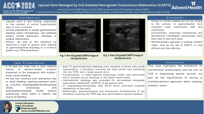
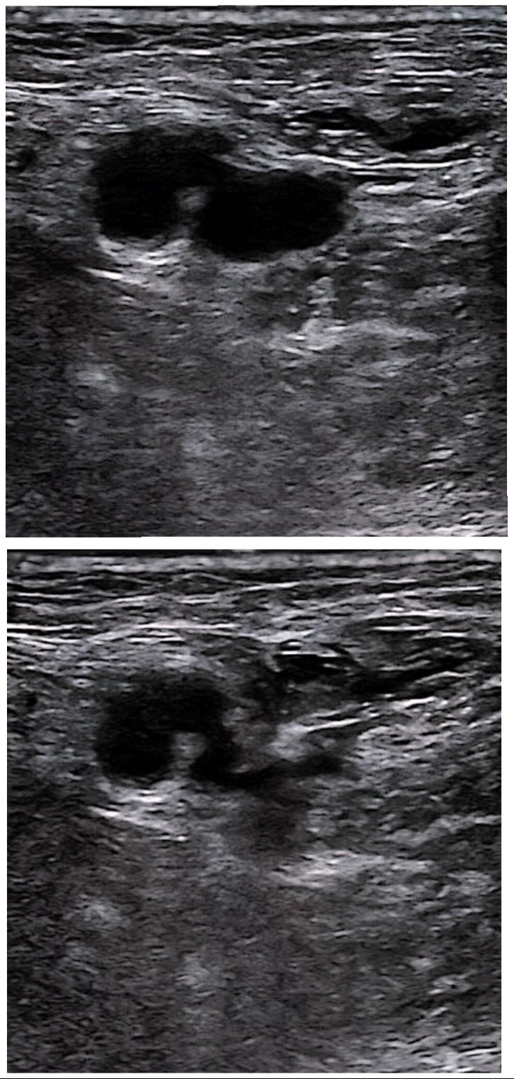
Figure: Pre and post US-guided CARTO pictures of jejunal varix
Disclosures:
Hamza Akram indicated no relevant financial relationships.
Qayum Shah indicated no relevant financial relationships.
Sanjeevani Tomar indicated no relevant financial relationships.
Qazi Wahab indicated no relevant financial relationships.
Hamza T. Akram, MBBS1, Qayum A. Shah, MBBS2, Sanjeevani Tomar, MD3, Qazi M.. Wahab, MBBS4. P2492 - Jejunal Varix Managed by Coil-Assisted Retrograde Transvenous Obliteration (CARTO): A Case Report, ACG 2024 Annual Scientific Meeting Abstracts. Philadelphia, PA: American College of Gastroenterology.
