Monday Poster Session
Category: GI Bleeding
P2493 - Splenic Artery Pseudoaneurysm Rupture in Gastric Remnant Presenting as Melena
Monday, October 28, 2024
10:30 AM - 4:00 PM ET
Location: Exhibit Hall E

Has Audio
- ES
Elen Sarkisyan, DO
Valley Hospital Medical Center
Las Vegas, NV
Presenting Author(s)
Elen Sarkisyan, DO, Scott Diamond, DO, Vishvinder Sharma, MD
Valley Hospital Medical Center, Las Vegas, NV
Introduction: Gastric remnant hemorrhage refers to bleeding in the bypassed portion of stomach in patients that have undergone gastrectomy procedures. Splenic artery pseudoaneurysm (SAP) is a rare life-threatening complication of gastric bypass procedures resulting in hemorrhage and can manifest as hemosuccus pancreaticus. In patients with Roux-en-Y gastric bypass (RYGB) surgery, the bypassed stomach is not visualized via conventional endoscopic approach.
Case Description/Methods: A 58-year-old female with chronic pancreatitis and RYGB 12 years prior presented with rectal bleeding. She underwent evaluation for melena a week prior with EGD with push enteroscopy and colonoscopy negative for bleeding. This admission, her blood pressure was 95/56 and hemoglobin was 3.9. She required massive transfusion with 8 units of red blood cells transfused and required vasopressor support.
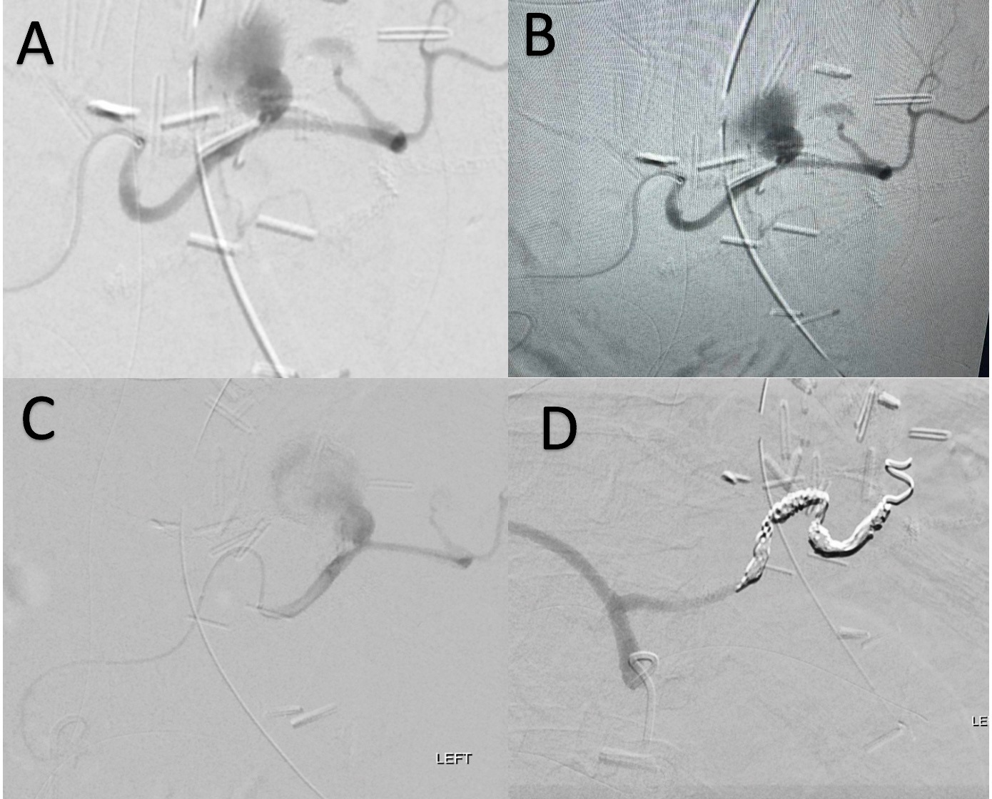
Computed tomography angiography (CTA) of the abdomen and pelvis was non-revealing. A nuclear medicine tagged RBC scan showed extravasated radioisotope in remnant stomach and small bowel. She was emergently taken for angiography by interventional radiology which revealed 2 large SAP with active contrast extravasation. The proximal SAP was successfully coil embolized with resolution of bleeding.
Discussion: SAP is a rare complication with 10 cases reported in an 18-year period and typically occurs as a result of weakening of the splenic artery. The weakened arterial wall of the SAP has potential to rupture and bleed through the ampulla of vater into the duodenum presenting as hemosuccus pancreaticus. SAP is the second most common cause of HP. RYGB anatomy limits endoscopic evaluation of the remnant stomach duodenum and pancreaticobiliary tree.
For evaluation of the bypassed gastric remnant and small intestine, more invasive options such as percutaneous distal gastrostomy and laparoscopic gastroduodenoscopy can be considered. Other tools include angiography, technetium-labeled RBC scan and celiac angiogram. Intermittent bleeding may not be captured with conventional CT imaging therefore physicians should consider CTA with delayed phase imaging or repeating technetium-labeled RBC scan. Angiography under fluoroscopy also provides an additional benefit of sequential imaging as compared to conventional CT.
With the rising prevalence of bariatric surgery, conventional endoscopic evaluation and treatment for upper GI bleeding becomes limited thus necessitating the use of dynamic imaging and interdisciplinary approach to improve patient outcomes.

Disclosures:
Elen Sarkisyan, DO, Scott Diamond, DO, Vishvinder Sharma, MD. P2493 - Splenic Artery Pseudoaneurysm Rupture in Gastric Remnant Presenting as Melena, ACG 2024 Annual Scientific Meeting Abstracts. Philadelphia, PA: American College of Gastroenterology.
Valley Hospital Medical Center, Las Vegas, NV
Introduction: Gastric remnant hemorrhage refers to bleeding in the bypassed portion of stomach in patients that have undergone gastrectomy procedures. Splenic artery pseudoaneurysm (SAP) is a rare life-threatening complication of gastric bypass procedures resulting in hemorrhage and can manifest as hemosuccus pancreaticus. In patients with Roux-en-Y gastric bypass (RYGB) surgery, the bypassed stomach is not visualized via conventional endoscopic approach.
Case Description/Methods: A 58-year-old female with chronic pancreatitis and RYGB 12 years prior presented with rectal bleeding. She underwent evaluation for melena a week prior with EGD with push enteroscopy and colonoscopy negative for bleeding. This admission, her blood pressure was 95/56 and hemoglobin was 3.9. She required massive transfusion with 8 units of red blood cells transfused and required vasopressor support.
Computed tomography angiography (CTA) of the abdomen and pelvis was non-revealing. A nuclear medicine tagged RBC scan showed extravasated radioisotope in remnant stomach and small bowel. She was emergently taken for angiography by interventional radiology which revealed 2 large SAP with active contrast extravasation. The proximal SAP was successfully coil embolized with resolution of bleeding.
Discussion: SAP is a rare complication with 10 cases reported in an 18-year period and typically occurs as a result of weakening of the splenic artery. The weakened arterial wall of the SAP has potential to rupture and bleed through the ampulla of vater into the duodenum presenting as hemosuccus pancreaticus. SAP is the second most common cause of HP. RYGB anatomy limits endoscopic evaluation of the remnant stomach duodenum and pancreaticobiliary tree.
For evaluation of the bypassed gastric remnant and small intestine, more invasive options such as percutaneous distal gastrostomy and laparoscopic gastroduodenoscopy can be considered. Other tools include angiography, technetium-labeled RBC scan and celiac angiogram. Intermittent bleeding may not be captured with conventional CT imaging therefore physicians should consider CTA with delayed phase imaging or repeating technetium-labeled RBC scan. Angiography under fluoroscopy also provides an additional benefit of sequential imaging as compared to conventional CT.
With the rising prevalence of bariatric surgery, conventional endoscopic evaluation and treatment for upper GI bleeding becomes limited thus necessitating the use of dynamic imaging and interdisciplinary approach to improve patient outcomes.

Figure: A, B, C) CT angiography reveals active extravasation from splenic artery
D) Splenic artery following successful coil embolization
D) Splenic artery following successful coil embolization
Disclosures:
Elen Sarkisyan indicated no relevant financial relationships.
Scott Diamond indicated no relevant financial relationships.
Vishvinder Sharma indicated no relevant financial relationships.
Elen Sarkisyan, DO, Scott Diamond, DO, Vishvinder Sharma, MD. P2493 - Splenic Artery Pseudoaneurysm Rupture in Gastric Remnant Presenting as Melena, ACG 2024 Annual Scientific Meeting Abstracts. Philadelphia, PA: American College of Gastroenterology.
