Monday Poster Session
Category: GI Bleeding
P2510 - The Uncommon Culprit: A Case of Upper GI Bleeding Secondary to an Ulcerated Gastric Lipoma
Monday, October 28, 2024
10:30 AM - 4:00 PM ET
Location: Exhibit Hall E

Has Audio

Brittney Shupp, DO
St. Luke's University Health Network
Bethlehem, PA
Presenting Author(s)
Brittney Shupp, DO1, Gurshawn Singh, MD2, Shannon Tosounian, DO1
1St. Luke's University Health Network, Bethlehem, PA; 2St. Luke's University Hospital, Bethlehem, PA
Introduction: Gastrointestinal (GI) lipomas are benign tumors that are often asymptomatic and incidentally found in the GI tract. Gastric lipomas are rare and account for only 1% of all stomach tumors but even more uncommonly lead to GI hemorrhage. We present a case of a GI bleed due to an ulcerated gastric lipoma.
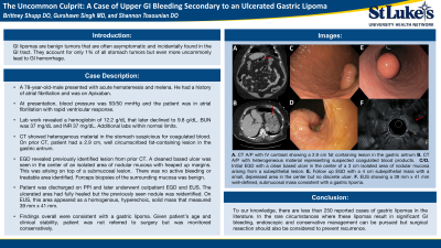
Case Description/Methods: A 78-year-old-male presented with acute hematemesis and melena. He had a history of atrial fibrillation and for this reason had been taking apixaban. At presentation, blood pressure was 93/50 mmHg and the patient was in atrial fibrillation with rapid ventricular response. Lab work revealed a hemoglobin of 12.2 g/dL that shortly after declined to 9.8 g/dL. Remaining lab work was unremarkable including INR of 1.15 seconds, but blood urea nitrogen (BUN) was 37 mg/dL. Computed tomography (CT) bleeding scan showed no active hemorrhage but heterogenous material seen in the stomach was suspicious for coagulated blood. On review of prior CT abdominal imaging, patient had a 2.9 cm, well circumscribed fat-containing lesion in the gastric antrum.
Patient underwent esophagogastroduodenoscopy (EGD) at which time the previously identified lesion from prior CT was seen. A cleaned based ulcer arose in the center of an isolated area of nodular mucosa with heaped up margins. This was arising on top of a submucosal lesion. There was no active bleeding or treatable area identified and the patient fortunately remained hemodynamically stable with no further overt bleeding. Forceps biopsies of the surrounding mucosa was benign. He was discharged on proton pump inhibitor therapy (PPI) but later underwent outpatient EGD and endoscopic ultrasound (EUS). The previously identified ulcerated area had fully healed. However the previously seen nodule was reidentified. On EUS, this area appeared as a homogenous, hyperechoic, solid mass that measured 39 mm by 41 mm. Findings overall were consistent with a gastric lipoma. Given patient’s age and clinical stability, patient was not referred to surgery but was monitored conservatively.
Discussion: To our knowledge, there are less than 250 reported cases of gastric lipomas in the literature. In the rare circumstances where these lipomas result in significant GI bleeding, endoscopic and conservative management can be pursued but surgical resection should also be considered to prevent recurrence. Additionally although uncommon, ulcerated lipomas should always be on the differential especially in patients with prior suggestive imaging.

Disclosures:
Brittney Shupp, DO1, Gurshawn Singh, MD2, Shannon Tosounian, DO1. P2510 - The Uncommon Culprit: A Case of Upper GI Bleeding Secondary to an Ulcerated Gastric Lipoma, ACG 2024 Annual Scientific Meeting Abstracts. Philadelphia, PA: American College of Gastroenterology.
1St. Luke's University Health Network, Bethlehem, PA; 2St. Luke's University Hospital, Bethlehem, PA
Introduction: Gastrointestinal (GI) lipomas are benign tumors that are often asymptomatic and incidentally found in the GI tract. Gastric lipomas are rare and account for only 1% of all stomach tumors but even more uncommonly lead to GI hemorrhage. We present a case of a GI bleed due to an ulcerated gastric lipoma.
Case Description/Methods: A 78-year-old-male presented with acute hematemesis and melena. He had a history of atrial fibrillation and for this reason had been taking apixaban. At presentation, blood pressure was 93/50 mmHg and the patient was in atrial fibrillation with rapid ventricular response. Lab work revealed a hemoglobin of 12.2 g/dL that shortly after declined to 9.8 g/dL. Remaining lab work was unremarkable including INR of 1.15 seconds, but blood urea nitrogen (BUN) was 37 mg/dL. Computed tomography (CT) bleeding scan showed no active hemorrhage but heterogenous material seen in the stomach was suspicious for coagulated blood. On review of prior CT abdominal imaging, patient had a 2.9 cm, well circumscribed fat-containing lesion in the gastric antrum.
Patient underwent esophagogastroduodenoscopy (EGD) at which time the previously identified lesion from prior CT was seen. A cleaned based ulcer arose in the center of an isolated area of nodular mucosa with heaped up margins. This was arising on top of a submucosal lesion. There was no active bleeding or treatable area identified and the patient fortunately remained hemodynamically stable with no further overt bleeding. Forceps biopsies of the surrounding mucosa was benign. He was discharged on proton pump inhibitor therapy (PPI) but later underwent outpatient EGD and endoscopic ultrasound (EUS). The previously identified ulcerated area had fully healed. However the previously seen nodule was reidentified. On EUS, this area appeared as a homogenous, hyperechoic, solid mass that measured 39 mm by 41 mm. Findings overall were consistent with a gastric lipoma. Given patient’s age and clinical stability, patient was not referred to surgery but was monitored conservatively.
Discussion: To our knowledge, there are less than 250 reported cases of gastric lipomas in the literature. In the rare circumstances where these lipomas result in significant GI bleeding, endoscopic and conservative management can be pursued but surgical resection should also be considered to prevent recurrence. Additionally although uncommon, ulcerated lipomas should always be on the differential especially in patients with prior suggestive imaging.

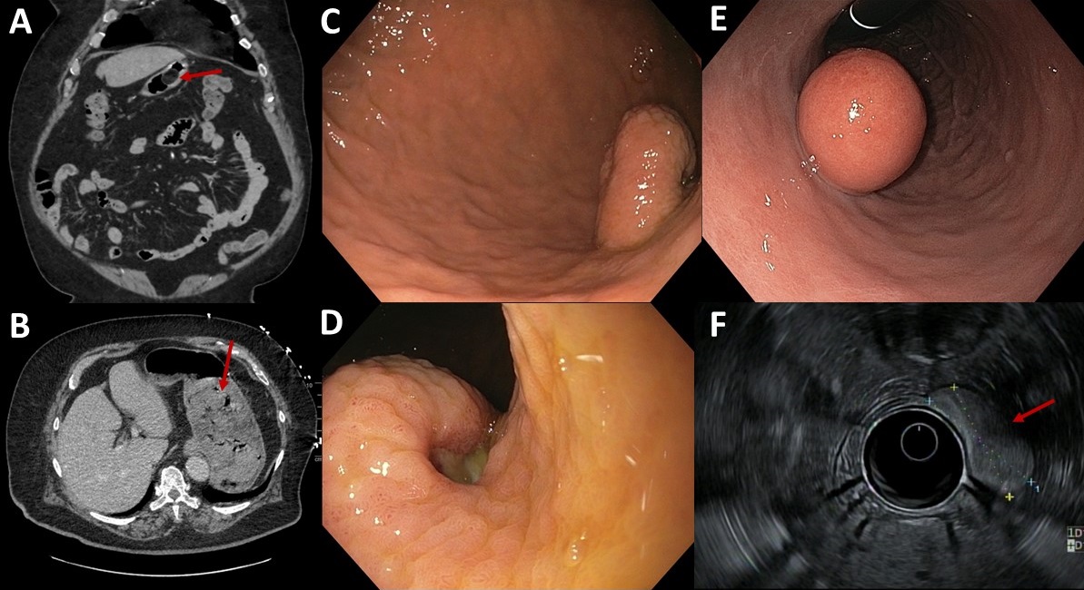
Figure: Image 1: A. CT abdomen pelvis with IV contrast which shows a well circumscribed, fat containing lesion in the gastric antrum measuring 2.9 cm. B. CT high volume bleeding scan completed on initial admission showing no evidence of active high volume gastrointestinal hemorrhage, but somewhat heterogeneous material seen throughout the stomach representing suspected coagulated blood products. C/D. EGD showing a healing, clean based ulcer in center of 3 cm isolated area of nodular mucosa with heaped up margins arising from a submucosal lesion. E. EGD showing a 4 cm subepithelial mass with a small, depressed area in the center but no discrete ulcer identified. F. EUS with a loculated, oval, homogenous, hyperechoic and solid mass measuring 39 mm x 41 mm with well-defined and smooth margins, contained within the submucosa consistent with a gastric lipoma.
Disclosures:
Brittney Shupp indicated no relevant financial relationships.
Gurshawn Singh indicated no relevant financial relationships.
Shannon Tosounian indicated no relevant financial relationships.
Brittney Shupp, DO1, Gurshawn Singh, MD2, Shannon Tosounian, DO1. P2510 - The Uncommon Culprit: A Case of Upper GI Bleeding Secondary to an Ulcerated Gastric Lipoma, ACG 2024 Annual Scientific Meeting Abstracts. Philadelphia, PA: American College of Gastroenterology.
