Monday Poster Session
Category: IBD
P2740 - Eye Opener: Orbital Myositis Unveiling Crohn's Disease
Monday, October 28, 2024
10:30 AM - 4:00 PM ET
Location: Exhibit Hall E

Has Audio
- CM
Cesar Moreno, MD
USA Health, University of South Alabama
Mobile, AL
Presenting Author(s)
Award: Presidential Poster Award
Elizabeth Statham, MD1, Cesar Moreno, MD2, Mary C. Marshall, MD3
1University of Alabama at Birmingham, Birmingham, AL; 2USA Health, University of South Alabama, Mobile, AL; 3University of South Alabama, Mobile, AL
Introduction: Orbital myositis (OM) is a rare cause of extraocular muscle inflammation. It can present as an extraintestinal manifestation of Crohn’s disease (CD). Ocular manifestations are present in about 4-10% of patients with inflammatory bowel disease (IBD), with the majority consisting of uveitis, scleritis, and episcleritis. We present the case of a 19-year-old female with previously undiagnosed Crohn’s disease who had presented with OM four years prior.
Case Description/Methods: A 19-year-old female was evaluated by ophthalmology four years prior due to ocular pain, diplopia, and periorbital edema. Workup revealed positive antinuclear antibodies 1:160, erythrocyte sedimentation rate of 53 mm/hr, normal thyroid function, and negative rheumatoid factor. Additional evaluation was negative for thyroid disease, sarcoidosis, and IgG4-related disease. She was diagnosed with OM after imaging showed enlargement and inflammation of extraocular muscles. Diplopia resolved after initiation of methotrexate and methylprednisolone. Over the next 4 years, she developed OM flares (figure 1) which were managed with steroid tapers, methotrexate, mycophenolate, and eventually rituximab. Four years after OM diagnosis, the patient developed oral ulcers and odynophagia requiring hospitalization. After discharge, the patient was referred to gastroenterology for evaluation of CD given mucositis and elevated fecal calprotectin. Endoscopy showed duodenitis, terminal ileitis, left sided colitis and rectal ulcers (figure 1). Biopsies were consistent with CD. The patient was started on adalimumab which led to clinical improvement and no subsequent OM flares.
Discussion: OM involves inflammation of one or more extraocular muscles. It can be caused by autoimmune conditions such as rheumatoid arthritis, sarcoidosis, systemic lupus erythematosus, and IBD. Clinical presentation may include orbital edema, proptosis, diplopia, conjunctival injection or ophthalmalgia. OM in patients with IBD may be due to a type III hypersensitivity reaction between colon proteins and extraocular muscles. OM can occur independent of gastrointestinal disease activity: it may occur in IBD clinical remission or years prior to development of gastrointestinal symptoms. This case emphasizes the importance of considering IBD in patients with ocular disease and systemic inflammation despite the lack of initial gastrointestinal symptoms.

Disclosures:
Elizabeth Statham, MD1, Cesar Moreno, MD2, Mary C. Marshall, MD3. P2740 - Eye Opener: Orbital Myositis Unveiling Crohn's Disease, ACG 2024 Annual Scientific Meeting Abstracts. Philadelphia, PA: American College of Gastroenterology.
Elizabeth Statham, MD1, Cesar Moreno, MD2, Mary C. Marshall, MD3
1University of Alabama at Birmingham, Birmingham, AL; 2USA Health, University of South Alabama, Mobile, AL; 3University of South Alabama, Mobile, AL
Introduction: Orbital myositis (OM) is a rare cause of extraocular muscle inflammation. It can present as an extraintestinal manifestation of Crohn’s disease (CD). Ocular manifestations are present in about 4-10% of patients with inflammatory bowel disease (IBD), with the majority consisting of uveitis, scleritis, and episcleritis. We present the case of a 19-year-old female with previously undiagnosed Crohn’s disease who had presented with OM four years prior.
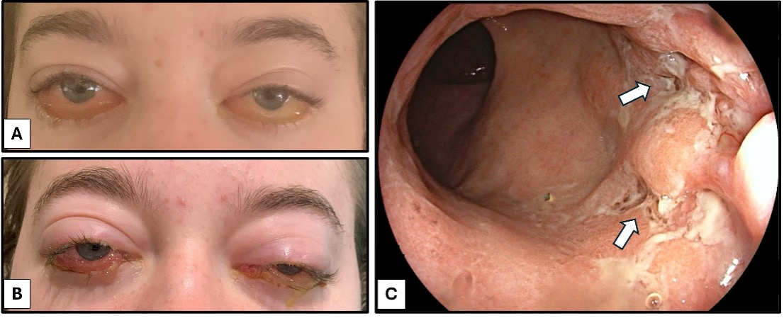
Case Description/Methods: A 19-year-old female was evaluated by ophthalmology four years prior due to ocular pain, diplopia, and periorbital edema. Workup revealed positive antinuclear antibodies 1:160, erythrocyte sedimentation rate of 53 mm/hr, normal thyroid function, and negative rheumatoid factor. Additional evaluation was negative for thyroid disease, sarcoidosis, and IgG4-related disease. She was diagnosed with OM after imaging showed enlargement and inflammation of extraocular muscles. Diplopia resolved after initiation of methotrexate and methylprednisolone. Over the next 4 years, she developed OM flares (figure 1) which were managed with steroid tapers, methotrexate, mycophenolate, and eventually rituximab. Four years after OM diagnosis, the patient developed oral ulcers and odynophagia requiring hospitalization. After discharge, the patient was referred to gastroenterology for evaluation of CD given mucositis and elevated fecal calprotectin. Endoscopy showed duodenitis, terminal ileitis, left sided colitis and rectal ulcers (figure 1). Biopsies were consistent with CD. The patient was started on adalimumab which led to clinical improvement and no subsequent OM flares.
Discussion: OM involves inflammation of one or more extraocular muscles. It can be caused by autoimmune conditions such as rheumatoid arthritis, sarcoidosis, systemic lupus erythematosus, and IBD. Clinical presentation may include orbital edema, proptosis, diplopia, conjunctival injection or ophthalmalgia. OM in patients with IBD may be due to a type III hypersensitivity reaction between colon proteins and extraocular muscles. OM can occur independent of gastrointestinal disease activity: it may occur in IBD clinical remission or years prior to development of gastrointestinal symptoms. This case emphasizes the importance of considering IBD in patients with ocular disease and systemic inflammation despite the lack of initial gastrointestinal symptoms.

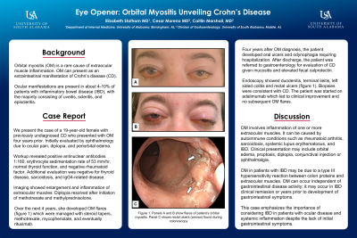
Figure: Figure 1. Panels A and B show flares of patient’s orbital myositis. Panel C shows rectal ulcers (arrows) found during colonoscopy
Disclosures:
Elizabeth Statham indicated no relevant financial relationships.
Cesar Moreno indicated no relevant financial relationships.
Mary Marshall indicated no relevant financial relationships.
Elizabeth Statham, MD1, Cesar Moreno, MD2, Mary C. Marshall, MD3. P2740 - Eye Opener: Orbital Myositis Unveiling Crohn's Disease, ACG 2024 Annual Scientific Meeting Abstracts. Philadelphia, PA: American College of Gastroenterology.

