Monday Poster Session
Category: Interventional Endoscopy
P2812 - An Unusual Presentation of Esophageal Adenocarcinoma Cured with Endoscopic Mucosal Resection
Monday, October 28, 2024
10:30 AM - 4:00 PM ET
Location: Exhibit Hall E

Has Audio
- CP
Chandler Patton, DO
Lehigh Valley Health Network
Allentown, PA
Presenting Author(s)
Chandler Patton, DO1, Shashin Shah, MD2, William Ghaul, DO1, Amanda Jacubowsky, DO1, Timothy Graziano, DO1
1Lehigh Valley Health Network, Allentown, PA; 2Eastern Pennsylvania Gastrointestinal and Liver Specialists, Allentown, PA
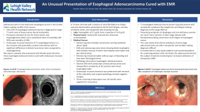
Introduction: Adenocarcinoma is the dominant esophageal cancer in the United States. It makes up around 60% of all esophageal cancers with 20% of the cases being diagnosed at stage T1. Previously the standard of care for treatment was surgical intervention with esophagectomy. Here we have a patient who presented with bloody stools and was found to have esophageal adenocarcinoma requiring endoscopic mucosal resection (EMR) for esophageal adenocarcinoma.
Case Description/Methods: A 73-year-old male with a history of atrial fibrillation on Eliquis presented to the hospital with concerns of fatigue, one day of bloody stool, and outpatient testing revealing hemoglobin of 5.2 g/dL (baseline 14.6 g/dL). On arrival, he was tachycardiac but otherwise vitally stable, and his laboratory testing was significant for a low MCV and a slightly elevated BUN. He was transfused and gastroenterology was consulted. An upper endoscopy and colonoscopy were performed and were significant for distal esophagitis with nodular mucosa of which two biopsies were taken. He remained stable and was discharged with outpatient follow-up. The patient’s biopsy results showed adenocarcinoma of the esophagus. Repeat upper endoscopy with endoscopic ultrasound showed area of nodularity at GE junction with intact esophageal walls without sign of adenocarcinoma invasion. Endoscopic mucosal resection was then performed with the removal of nodularity. Pathology revealed T1a esophageal adenocarcinoma with negative margins. The patient had screening EGD’s with a biopsy of the resection site every 6 months for three occurrences which showed no recurrence of adenocarcinoma at the resection site.
Discussion: This case discusses a rare presentation of hematochezia resulting in the diagnosis of T1a esophageal carcinoma that typically presents with weight loss and fatigue with more rare symptoms being dysphasia and iron deficiency anemia. The later symptoms typically occur in more advanced stages of the disease with only 25% of patients having localized disease on presentation with very few cases reporting overt hematochezia. This case demonstrates the importance of biopsy of nodular esophageal mucosa even during endoscopic evaluation of hematochezia given the potential for curative intervention via EMR in the setting of T1 adenocarcinoma.

Disclosures:
Chandler Patton, DO1, Shashin Shah, MD2, William Ghaul, DO1, Amanda Jacubowsky, DO1, Timothy Graziano, DO1. P2812 - An Unusual Presentation of Esophageal Adenocarcinoma Cured with Endoscopic Mucosal Resection, ACG 2024 Annual Scientific Meeting Abstracts. Philadelphia, PA: American College of Gastroenterology.
1Lehigh Valley Health Network, Allentown, PA; 2Eastern Pennsylvania Gastrointestinal and Liver Specialists, Allentown, PA
Introduction: Adenocarcinoma is the dominant esophageal cancer in the United States. It makes up around 60% of all esophageal cancers with 20% of the cases being diagnosed at stage T1. Previously the standard of care for treatment was surgical intervention with esophagectomy. Here we have a patient who presented with bloody stools and was found to have esophageal adenocarcinoma requiring endoscopic mucosal resection (EMR) for esophageal adenocarcinoma.
Case Description/Methods: A 73-year-old male with a history of atrial fibrillation on Eliquis presented to the hospital with concerns of fatigue, one day of bloody stool, and outpatient testing revealing hemoglobin of 5.2 g/dL (baseline 14.6 g/dL). On arrival, he was tachycardiac but otherwise vitally stable, and his laboratory testing was significant for a low MCV and a slightly elevated BUN. He was transfused and gastroenterology was consulted. An upper endoscopy and colonoscopy were performed and were significant for distal esophagitis with nodular mucosa of which two biopsies were taken. He remained stable and was discharged with outpatient follow-up. The patient’s biopsy results showed adenocarcinoma of the esophagus. Repeat upper endoscopy with endoscopic ultrasound showed area of nodularity at GE junction with intact esophageal walls without sign of adenocarcinoma invasion. Endoscopic mucosal resection was then performed with the removal of nodularity. Pathology revealed T1a esophageal adenocarcinoma with negative margins. The patient had screening EGD’s with a biopsy of the resection site every 6 months for three occurrences which showed no recurrence of adenocarcinoma at the resection site.
Discussion: This case discusses a rare presentation of hematochezia resulting in the diagnosis of T1a esophageal carcinoma that typically presents with weight loss and fatigue with more rare symptoms being dysphasia and iron deficiency anemia. The later symptoms typically occur in more advanced stages of the disease with only 25% of patients having localized disease on presentation with very few cases reporting overt hematochezia. This case demonstrates the importance of biopsy of nodular esophageal mucosa even during endoscopic evaluation of hematochezia given the potential for curative intervention via EMR in the setting of T1 adenocarcinoma.

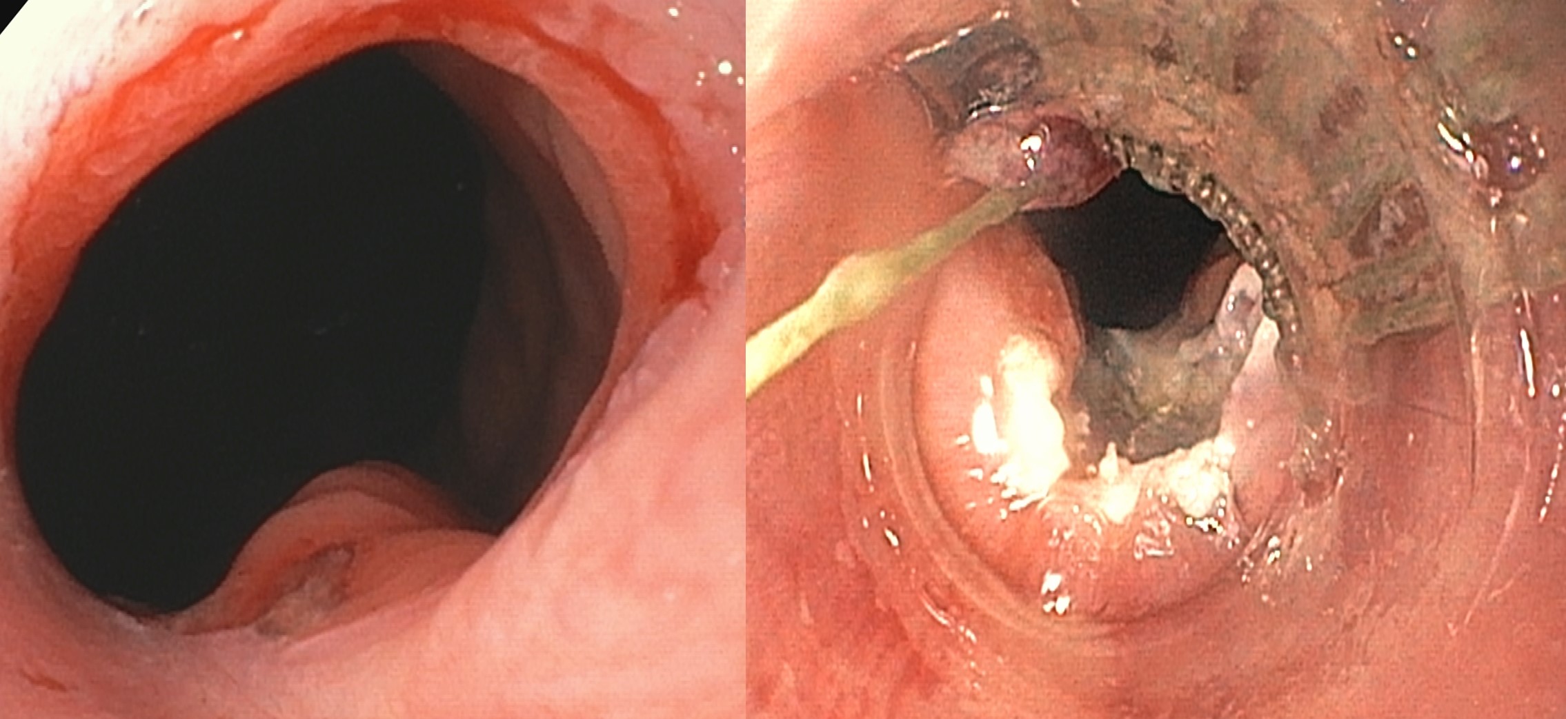
Figure: Figures A and B: Esophageal adenocarcinoma before and after completion of endoscopic mucosal resection
Disclosures:
Chandler Patton indicated no relevant financial relationships.
Shashin Shah indicated no relevant financial relationships.
William Ghaul indicated no relevant financial relationships.
Amanda Jacubowsky indicated no relevant financial relationships.
Timothy Graziano indicated no relevant financial relationships.
Chandler Patton, DO1, Shashin Shah, MD2, William Ghaul, DO1, Amanda Jacubowsky, DO1, Timothy Graziano, DO1. P2812 - An Unusual Presentation of Esophageal Adenocarcinoma Cured with Endoscopic Mucosal Resection, ACG 2024 Annual Scientific Meeting Abstracts. Philadelphia, PA: American College of Gastroenterology.
