Monday Poster Session
Category: Interventional Endoscopy
P2828 - How Large Can a Gallstone get? A Rare Case of Bouveret's Syndrome Treated With Electrohydraulic Lithotripsy and Mechanical Extraction
Monday, October 28, 2024
10:30 AM - 4:00 PM ET
Location: Exhibit Hall E

Has Audio

Kais Antonios, MD
Trinity Health Ann Arbor Hospital
Ann Arbor, MI
Presenting Author(s)
Kais Antonios, MD1, Sarvani Surapaneni, MD2, Michael Zijlstra, DO2, Wael Al-Yaman, MD2, Mohannad Abousaleh, MD3, Naresh Gunaratnam, MD4
1Trinity Health Ann Arbor Hospital, Ann Arbor, MI; 2Trinity Health Ann Arbor Hospital, Ypsilanti, MI; 3Ypsilanti, MI; 4Huron Gastro, Ann Arbor, MI
Introduction: Bouveret syndrome is defined as a gastric outlet obstruction that arises from gallstones impacted in the distal stomach or proximal duodenum after passing through a cholecystoduodenal, cholecystogastric or a choledocoduodenal fistula. It is responsible for 1-3 % of cases of gallstone ileus, and despite multiple endoscopic treatment options, these are successful in only one-third of the cases. Here, we describe a case of a patient with a 3.9 cm gallstone causing Bouveret syndrome that was successfully treated with an endoscopic approach.
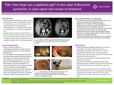
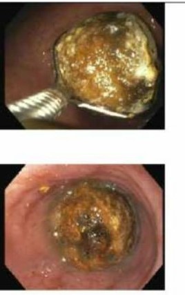
Case Description/Methods: An 82 yo male with a history of Cholecystoduodenal fistula resulting in chronic cholecystitis presented due to acute epigastric abdominal pain with CT of the abdomen demonstrating pneumobilia and inflammatory changes at the proximal part of the duodenum and the head of the pancreas. He was hemodynamically stable. Labs showed a leukocytosis of 18.1 K, and a lipase of 1636 (Reference range 11-82 U/L). After initiation of IV fluids and antibiotics, he underwent and EGD, in which a stone measuring >30 mm was found in the duodenal bulb obstructing the duodenum, and after mechanical and electrohydraulic lithotripsy were used, the stone was successfully broken down into smaller stones but unable to be completely removed. The patient had significant symptomatic improvement, and another attempt was performed the next day, and despite the use of many modalities including an electrohydraulic instrument, and a mechanical lithotripter, the stone removal was not completely successful. A 3rd EGD was carried on two days after, and after the bulb was flooded with normal saline, 6,316 shocks were delivered at high power, using 5 electrohydraulic probes which led to successful fragmentation and later removal of the stone in 4 separate fragments. Following the procedure, the patient had an uncomplicated course, and was discharged a day later from the hospital.
Discussion: Multiple enoscopic modalities including stone removal or fragmentation with classical endoscopic devices, or fragmentation of gallstones with new devices, such as electrohydraulic lithotripsy, laser, and extracorporeal shockwave lithotripsy have been described in the endoscopic treatment of Bouveret syndrome. There has been no comparison of efficacy between these approaches in the literature. Our case demonstrates that combining electrohydraulic lithotripsy with other modalities lead to successful endoscopic treatment of Bouveret syndrome caused by a very large stone.

Disclosures:
Kais Antonios, MD1, Sarvani Surapaneni, MD2, Michael Zijlstra, DO2, Wael Al-Yaman, MD2, Mohannad Abousaleh, MD3, Naresh Gunaratnam, MD4. P2828 - How Large Can a Gallstone get? A Rare Case of Bouveret's Syndrome Treated With Electrohydraulic Lithotripsy and Mechanical Extraction, ACG 2024 Annual Scientific Meeting Abstracts. Philadelphia, PA: American College of Gastroenterology.
1Trinity Health Ann Arbor Hospital, Ann Arbor, MI; 2Trinity Health Ann Arbor Hospital, Ypsilanti, MI; 3Ypsilanti, MI; 4Huron Gastro, Ann Arbor, MI
Introduction: Bouveret syndrome is defined as a gastric outlet obstruction that arises from gallstones impacted in the distal stomach or proximal duodenum after passing through a cholecystoduodenal, cholecystogastric or a choledocoduodenal fistula. It is responsible for 1-3 % of cases of gallstone ileus, and despite multiple endoscopic treatment options, these are successful in only one-third of the cases. Here, we describe a case of a patient with a 3.9 cm gallstone causing Bouveret syndrome that was successfully treated with an endoscopic approach.
Case Description/Methods: An 82 yo male with a history of Cholecystoduodenal fistula resulting in chronic cholecystitis presented due to acute epigastric abdominal pain with CT of the abdomen demonstrating pneumobilia and inflammatory changes at the proximal part of the duodenum and the head of the pancreas. He was hemodynamically stable. Labs showed a leukocytosis of 18.1 K, and a lipase of 1636 (Reference range 11-82 U/L). After initiation of IV fluids and antibiotics, he underwent and EGD, in which a stone measuring >30 mm was found in the duodenal bulb obstructing the duodenum, and after mechanical and electrohydraulic lithotripsy were used, the stone was successfully broken down into smaller stones but unable to be completely removed. The patient had significant symptomatic improvement, and another attempt was performed the next day, and despite the use of many modalities including an electrohydraulic instrument, and a mechanical lithotripter, the stone removal was not completely successful. A 3rd EGD was carried on two days after, and after the bulb was flooded with normal saline, 6,316 shocks were delivered at high power, using 5 electrohydraulic probes which led to successful fragmentation and later removal of the stone in 4 separate fragments. Following the procedure, the patient had an uncomplicated course, and was discharged a day later from the hospital.
Discussion: Multiple enoscopic modalities including stone removal or fragmentation with classical endoscopic devices, or fragmentation of gallstones with new devices, such as electrohydraulic lithotripsy, laser, and extracorporeal shockwave lithotripsy have been described in the endoscopic treatment of Bouveret syndrome. There has been no comparison of efficacy between these approaches in the literature. Our case demonstrates that combining electrohydraulic lithotripsy with other modalities lead to successful endoscopic treatment of Bouveret syndrome caused by a very large stone.

Figure: EGD showing a large gallstone causing duodenal obstruction with attempts of mechanical extraction
Disclosures:
Kais Antonios indicated no relevant financial relationships.
Sarvani Surapaneni indicated no relevant financial relationships.
Michael Zijlstra indicated no relevant financial relationships.
Wael Al-Yaman indicated no relevant financial relationships.
Mohannad Abousaleh indicated no relevant financial relationships.
Naresh Gunaratnam indicated no relevant financial relationships.
Kais Antonios, MD1, Sarvani Surapaneni, MD2, Michael Zijlstra, DO2, Wael Al-Yaman, MD2, Mohannad Abousaleh, MD3, Naresh Gunaratnam, MD4. P2828 - How Large Can a Gallstone get? A Rare Case of Bouveret's Syndrome Treated With Electrohydraulic Lithotripsy and Mechanical Extraction, ACG 2024 Annual Scientific Meeting Abstracts. Philadelphia, PA: American College of Gastroenterology.
