Monday Poster Session
Category: Interventional Endoscopy
P2830 - Endoscopic Ultrasound-Guided Gastric Remnant Access in a Patient With Massive Upper Gastrointestinal Bleeding and a History of a Roux-en-Y Gastric Bypass After Failed IR Embolization of a Bleeding Duodenal Ulcer
Monday, October 28, 2024
10:30 AM - 4:00 PM ET
Location: Exhibit Hall E

Has Audio

Jessica Basso, MD
Brooke Army Medical Center
Fort Sam Houston, TX
Presenting Author(s)
Award: Presidential Poster Award
Jessica Basso, MD, Jerome C. Edelson, MD
Brooke Army Medical Center, Fort Sam Houston, TX
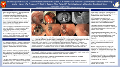
Introduction: Gastric remnant bleeding is a rare complication of Roux-en-Y Gastric Bypass (RYGB). It is hypothesized that altered pathophysiologic environment of excluded anatomy and bile reflux can result in gastritis and peptic ulcer disease. Conventional endoscopic techniques to access the remnant include single balloon enteroscopy and laparoscopic-assisted endoscopy. Endoscopic Ultrasound (EUS)-directed transgastric intervention (EDGI), is a newer endoscopic option that utilizes a lumen-apposing metal stent (LAMS) to facilitate access into the gastric remnant. We present a case of acute GI bleeding in the duodenum after RYGB, managed with an over the scope clip placed during a single-session EDGI.
Case Description/Methods: A 72-year-old female with RYGB and breast cancer on trastuzumab presented with abdominal pain and melena. Upper endoscopy revealed a marginal ulcer (Figure 1A) without high-risk stigmata. She returned with ongoing melena and worsening anemia. Repeat upper endoscopy showed bright red blood at the jejuno-jejunostomy (B) and single balloon enteroscopy did not identify etiology for bleeding in the roux limb. A CT Angiography (C) revealed contrast extravasation in the duodenal bulb, although Interventional Radiology did not identify a bleeding source on abdominal arteriogram. The patient continued to have active bleeding, requiring blood transfusions. Following a multi-disciplinary discussion, the decision was made to pursue EDGI. The remnant stomach was located under EUS and a 10 mm x 20 mm hot LAMS was placed to create a gastrogastrostomy, secured with two sutures (D-E). A 15mm Forrest IIa ulcer was found on the anterior wall of the duodenum (F). An over the scope clip was deployed over the ulcer and epinephrine was injected around the clip (G). The patient’s condition improved and she was discharged. Repeat upper endoscopy 8 weeks later showed a healed duodenal ulcer with migration of the clip (H). The LAMS was removed.
Discussion: EGDI is developing as the preferred approach to enable access to the gastric remnant in patients with altered anatomy at centers with LAMS experience. It provides the ability to perform endoscopic interventions with higher technical success and fewer complications compared to traditional methods. This case highlights a minimally invasive approach to immediate diagnosis and management of bleeding in excluded anatomy through a single-session EDGI procedure. Further improvements in strategies and methods with this technique may reduce risks and improve outcomes.

Disclosures:
Jessica Basso, MD, Jerome C. Edelson, MD. P2830 - Endoscopic Ultrasound-Guided Gastric Remnant Access in a Patient With Massive Upper Gastrointestinal Bleeding and a History of a Roux-en-Y Gastric Bypass After Failed IR Embolization of a Bleeding Duodenal Ulcer, ACG 2024 Annual Scientific Meeting Abstracts. Philadelphia, PA: American College of Gastroenterology.
Jessica Basso, MD, Jerome C. Edelson, MD
Brooke Army Medical Center, Fort Sam Houston, TX
Introduction: Gastric remnant bleeding is a rare complication of Roux-en-Y Gastric Bypass (RYGB). It is hypothesized that altered pathophysiologic environment of excluded anatomy and bile reflux can result in gastritis and peptic ulcer disease. Conventional endoscopic techniques to access the remnant include single balloon enteroscopy and laparoscopic-assisted endoscopy. Endoscopic Ultrasound (EUS)-directed transgastric intervention (EDGI), is a newer endoscopic option that utilizes a lumen-apposing metal stent (LAMS) to facilitate access into the gastric remnant. We present a case of acute GI bleeding in the duodenum after RYGB, managed with an over the scope clip placed during a single-session EDGI.
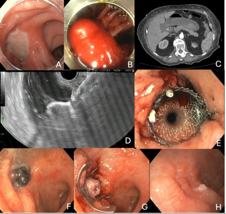
Case Description/Methods: A 72-year-old female with RYGB and breast cancer on trastuzumab presented with abdominal pain and melena. Upper endoscopy revealed a marginal ulcer (Figure 1A) without high-risk stigmata. She returned with ongoing melena and worsening anemia. Repeat upper endoscopy showed bright red blood at the jejuno-jejunostomy (B) and single balloon enteroscopy did not identify etiology for bleeding in the roux limb. A CT Angiography (C) revealed contrast extravasation in the duodenal bulb, although Interventional Radiology did not identify a bleeding source on abdominal arteriogram. The patient continued to have active bleeding, requiring blood transfusions. Following a multi-disciplinary discussion, the decision was made to pursue EDGI. The remnant stomach was located under EUS and a 10 mm x 20 mm hot LAMS was placed to create a gastrogastrostomy, secured with two sutures (D-E). A 15mm Forrest IIa ulcer was found on the anterior wall of the duodenum (F). An over the scope clip was deployed over the ulcer and epinephrine was injected around the clip (G). The patient’s condition improved and she was discharged. Repeat upper endoscopy 8 weeks later showed a healed duodenal ulcer with migration of the clip (H). The LAMS was removed.
Discussion: EGDI is developing as the preferred approach to enable access to the gastric remnant in patients with altered anatomy at centers with LAMS experience. It provides the ability to perform endoscopic interventions with higher technical success and fewer complications compared to traditional methods. This case highlights a minimally invasive approach to immediate diagnosis and management of bleeding in excluded anatomy through a single-session EDGI procedure. Further improvements in strategies and methods with this technique may reduce risks and improve outcomes.

Figure: (A) A 4cm clean based gastric ulcer at the GJ anastomosis
(B) Bright red blood and clot at the J-J anastomosis
(C) CT Abdomen and Pelvis with and without contrast showing a focus of contrast extravasation in the lumen of the 2nd portion of the
duodenum, which expands slightly on delayed imaging
(D) EUS guided electrocautery enhanced (hot) 10 x 20 mm LAMS deployed using autocut settings
(E) 2T endoscope overstitch system used for placement of two sutures
(F) A 15mm duodenum bulb ulcer with a large visible vessel (Forrest IIa) on the anterior wall
(G) An Over the Scope Clip deployed over the ulcer with 2.5 ml of epinephrine injected in 4 quadrants around the clip
(H) Healed duodenal ulcer with migration of the clip
Disclosures:
Jessica Basso indicated no relevant financial relationships.
Jerome Edelson indicated no relevant financial relationships.
Jessica Basso, MD, Jerome C. Edelson, MD. P2830 - Endoscopic Ultrasound-Guided Gastric Remnant Access in a Patient With Massive Upper Gastrointestinal Bleeding and a History of a Roux-en-Y Gastric Bypass After Failed IR Embolization of a Bleeding Duodenal Ulcer, ACG 2024 Annual Scientific Meeting Abstracts. Philadelphia, PA: American College of Gastroenterology.


