Monday Poster Session
Category: Interventional Endoscopy
P2833 - Combined Endovascular and Endoscopic Removal of a Migrated IVC Filter With Closure of Duodenal Fistula
Monday, October 28, 2024
10:30 AM - 4:00 PM ET
Location: Exhibit Hall E

Has Audio

Chandler Shapiro, MD
Rush University Medical Center
Chicago, IL
Presenting Author(s)
Chandler Shapiro, MD, Brittany Doll, MD, Zoë Post, MD, Christopher Chapman, MD
Rush University Medical Center, Chicago, IL
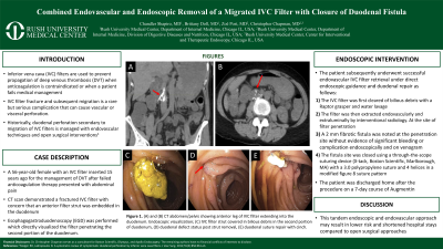
Introduction: Inferior vena cava (IVC) filters are used to prevent propagation of deep venous thrombosis (DVT) when anticoagulation is contraindicated or when a patient fails medical management. IVC filter fracture and subsequent migration is a rare but serious complication that can cause vascular or visceral perforation. Historically, duodenal perforation secondary to migration of IVC filters is managed with endovascular techniques and surgical interventions. Here we present a case of combined interventional radiology and endoscopic management of IVC filter migration with duodenal fistula repair.
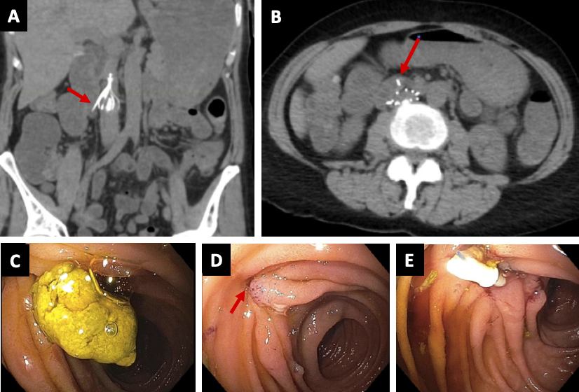
Case Description/Methods: A 56-year-old female with an IVC filter inserted 15 years ago for the management of DVT after failed anticoagulation therapy presented with abdominal pain. A computed tomography (CT) scan demonstrated a fractured IVC filter with concern that an anterior filter strut was embedded in the duodenum (figure 1A-B). Esophagogastroduodenoscopy (EGD) was performed which directly visualized the filter penetrating the second portion of the duodenum. The patient subsequently underwent successful endovascular IVC filter retrieval under direct endoscopic guidance and duodenal repair as follows: the IVC filter was first cleared of bilious debris with a Raptor grasper and water lavage endoscopically (figure 1C). The filter was then extracted endovascularly and extraluminally by interventional radiology. At the site of filter penetration, a small 2 mm fibrotic appearing fistula was present (figure 1D). No significant bleeding or complication was noted endoscopically and a venogram was performed without evidence of extravasation. To reduce the risk of a delayed complication, closure of the fistula site was achieved using a through-the-scope suturing device (X-tack, Boston Scientific, Marlborough, MA) (figure 1E.). A single X-tack device with 3.0 proline suture and 4 helices was used to close the fistula in a modified figure 8 suture pattern. The patient was discharged home after the procedure on a 7-day course of amoxicillin-clavulanate.
Discussion: Here we report a successful case of migrated IVC filter removal and duodenal fistula repair under direct endoscopic visualization with through-the-scope endoscopic suturing. Historically, high risk exploratory laparotomies have been performed for IVC filter retrieval and duodenal repair in cases of duodenal penetration. This tandem endoscopic and endovascular approach may result in lower risk and shortened hospital stays compared to surgical approaches.

Disclosures:
Chandler Shapiro, MD, Brittany Doll, MD, Zoë Post, MD, Christopher Chapman, MD. P2833 - Combined Endovascular and Endoscopic Removal of a Migrated IVC Filter With Closure of Duodenal Fistula, ACG 2024 Annual Scientific Meeting Abstracts. Philadelphia, PA: American College of Gastroenterology.
Rush University Medical Center, Chicago, IL
Introduction: Inferior vena cava (IVC) filters are used to prevent propagation of deep venous thrombosis (DVT) when anticoagulation is contraindicated or when a patient fails medical management. IVC filter fracture and subsequent migration is a rare but serious complication that can cause vascular or visceral perforation. Historically, duodenal perforation secondary to migration of IVC filters is managed with endovascular techniques and surgical interventions. Here we present a case of combined interventional radiology and endoscopic management of IVC filter migration with duodenal fistula repair.
Case Description/Methods: A 56-year-old female with an IVC filter inserted 15 years ago for the management of DVT after failed anticoagulation therapy presented with abdominal pain. A computed tomography (CT) scan demonstrated a fractured IVC filter with concern that an anterior filter strut was embedded in the duodenum (figure 1A-B). Esophagogastroduodenoscopy (EGD) was performed which directly visualized the filter penetrating the second portion of the duodenum. The patient subsequently underwent successful endovascular IVC filter retrieval under direct endoscopic guidance and duodenal repair as follows: the IVC filter was first cleared of bilious debris with a Raptor grasper and water lavage endoscopically (figure 1C). The filter was then extracted endovascularly and extraluminally by interventional radiology. At the site of filter penetration, a small 2 mm fibrotic appearing fistula was present (figure 1D). No significant bleeding or complication was noted endoscopically and a venogram was performed without evidence of extravasation. To reduce the risk of a delayed complication, closure of the fistula site was achieved using a through-the-scope suturing device (X-tack, Boston Scientific, Marlborough, MA) (figure 1E.). A single X-tack device with 3.0 proline suture and 4 helices was used to close the fistula in a modified figure 8 suture pattern. The patient was discharged home after the procedure on a 7-day course of amoxicillin-clavulanate.
Discussion: Here we report a successful case of migrated IVC filter removal and duodenal fistula repair under direct endoscopic visualization with through-the-scope endoscopic suturing. Historically, high risk exploratory laparotomies have been performed for IVC filter retrieval and duodenal repair in cases of duodenal penetration. This tandem endoscopic and endovascular approach may result in lower risk and shortened hospital stays compared to surgical approaches.

Figure: Figure 1. (A) and (B) CT abdomen/pelvis showing anterior leg of IVC filter extending into the duodenum. Endoscopic visualization; (C) IVC filter strut covered in bilious debris in the second portion of duodenum, (D) duodenal defect status post strut removal, (E) duodenal suture repair with cinch.
Disclosures:
Chandler Shapiro indicated no relevant financial relationships.
Brittany Doll indicated no relevant financial relationships.
Zoë Post indicated no relevant financial relationships.
Christopher Chapman: Apollo Endosurgery – Consultant. Boston Scientific – Consultant. Olympus – Consultant.
Chandler Shapiro, MD, Brittany Doll, MD, Zoë Post, MD, Christopher Chapman, MD. P2833 - Combined Endovascular and Endoscopic Removal of a Migrated IVC Filter With Closure of Duodenal Fistula, ACG 2024 Annual Scientific Meeting Abstracts. Philadelphia, PA: American College of Gastroenterology.
