Monday Poster Session
Category: Interventional Endoscopy
P2845 - Duodenal Diverticulum Posing as Diffuse Large B-Cell Lymphoma
Monday, October 28, 2024
10:30 AM - 4:00 PM ET
Location: Exhibit Hall E

Has Audio
- AT
Alexander J. Thomas, MD
SSM Health Saint Louis University Hospital
St. Louis, MO
Presenting Author(s)
Alexander J. Thomas, MD1, Radhika Patel, MD1, Pradeep Yarra, MD2, Shravya Ginnaram, MD3, Marina Kim, DO1
1SSM Health Saint Louis University Hospital, St. Louis, MO; 2Saint Louis University School of Medicine, St. Louis, MO; 3Jefferson Abington Hospital, Willow Grove, PA
Introduction: Lymphoma is a heterogeneous group of malignancies which arise from lymphocytes. The majority of gastrointestinal (GI) lymphomas are extra nodal marginal zone B cell lymphoma of mucosa-associated lymphoid tissue (MALT) or Diffuse Large B-Cell Lymphoma (DLBCL). DLBCL typically presents with nodal enlargement in the neck or abdomen. We report an elderly woman presenting with syncope and unusual findings on cross-sectional imaging that eventually led to the diagnosis of DLBCL.
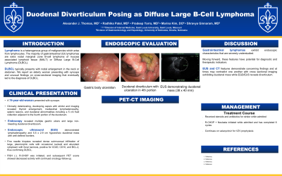
Case Description/Methods: A 74-year-old woman who presented with syncope, however initial workup was unrevealing. Clinically deteriorating, she developed sepsis with stridor, prompting further imaging with computed tomography (CT) of her chest, abdomen, and pelvis which revealed thyroid enlargement, mediastinal lymphadenopathy, splenic lesions, and duodenal abnormalities including a 5 cm fluid collection adjacent to the fourth portion of the duodenum. Endoscopy revealed multiple gastric ulcers and large non-bleeding duodenal diverticulum, in the fourth portion of the duodenum associated with an adjacent large, 3 cm duodenal ulcer. Further evaluation with endoscopic ultrasound (EUS demonstrated widespread lymphadenopathy and 5.5 x 2.9 cm hypoechoic duodenal mass with well-defined borders. Fine needle biopsies revealed dense submucosal infiltration of large, pleomorphic cells with occasional nucleoli and abundant cytoplasm with focal necrosis, positive for CD20, CD10, and BCL-2, thus confirming DLBCL. FISH was negative. R-CHOP chemotherapy was initiated while inpatient. Subsequent PET scans showed decreased activity with continued oncology follow-up.
Discussion: Rare gastrointestinal lymphomas, whether primary or secondary, exhibit understudied endoscopic characteristics. However, these features are gaining recognition as potential prognostic and therapeutic indicators. This case highlights an uncommon presentation that posed a diagnostic challenge. Diagnosis is made via endoscopy showing polyploid mass lesion, gastric fold thickening, gastric/peptic ulcer, or mucosal erythema. Concerns for disease may also be visualized on imaging, as our case demonstrated was visible on CT scan or EUS. The presence of a large diverticulum, especially in an unusual location, such as beyond the second portion of the duodenum, can sometimes mimic a mass on cross-sectional imaging. Treatment typically involves chemotherapy, as in our patient with R-CHOP therapy but early suspicion and diagnosis is paramount.
Disclosures:
Alexander J. Thomas, MD1, Radhika Patel, MD1, Pradeep Yarra, MD2, Shravya Ginnaram, MD3, Marina Kim, DO1. P2845 - Duodenal Diverticulum Posing as Diffuse Large B-Cell Lymphoma, ACG 2024 Annual Scientific Meeting Abstracts. Philadelphia, PA: American College of Gastroenterology.
1SSM Health Saint Louis University Hospital, St. Louis, MO; 2Saint Louis University School of Medicine, St. Louis, MO; 3Jefferson Abington Hospital, Willow Grove, PA
Introduction: Lymphoma is a heterogeneous group of malignancies which arise from lymphocytes. The majority of gastrointestinal (GI) lymphomas are extra nodal marginal zone B cell lymphoma of mucosa-associated lymphoid tissue (MALT) or Diffuse Large B-Cell Lymphoma (DLBCL). DLBCL typically presents with nodal enlargement in the neck or abdomen. We report an elderly woman presenting with syncope and unusual findings on cross-sectional imaging that eventually led to the diagnosis of DLBCL.
Case Description/Methods: A 74-year-old woman who presented with syncope, however initial workup was unrevealing. Clinically deteriorating, she developed sepsis with stridor, prompting further imaging with computed tomography (CT) of her chest, abdomen, and pelvis which revealed thyroid enlargement, mediastinal lymphadenopathy, splenic lesions, and duodenal abnormalities including a 5 cm fluid collection adjacent to the fourth portion of the duodenum. Endoscopy revealed multiple gastric ulcers and large non-bleeding duodenal diverticulum, in the fourth portion of the duodenum associated with an adjacent large, 3 cm duodenal ulcer. Further evaluation with endoscopic ultrasound (EUS demonstrated widespread lymphadenopathy and 5.5 x 2.9 cm hypoechoic duodenal mass with well-defined borders. Fine needle biopsies revealed dense submucosal infiltration of large, pleomorphic cells with occasional nucleoli and abundant cytoplasm with focal necrosis, positive for CD20, CD10, and BCL-2, thus confirming DLBCL. FISH was negative. R-CHOP chemotherapy was initiated while inpatient. Subsequent PET scans showed decreased activity with continued oncology follow-up.
Discussion: Rare gastrointestinal lymphomas, whether primary or secondary, exhibit understudied endoscopic characteristics. However, these features are gaining recognition as potential prognostic and therapeutic indicators. This case highlights an uncommon presentation that posed a diagnostic challenge. Diagnosis is made via endoscopy showing polyploid mass lesion, gastric fold thickening, gastric/peptic ulcer, or mucosal erythema. Concerns for disease may also be visualized on imaging, as our case demonstrated was visible on CT scan or EUS. The presence of a large diverticulum, especially in an unusual location, such as beyond the second portion of the duodenum, can sometimes mimic a mass on cross-sectional imaging. Treatment typically involves chemotherapy, as in our patient with R-CHOP therapy but early suspicion and diagnosis is paramount.
Disclosures:
Alexander Thomas indicated no relevant financial relationships.
Radhika Patel indicated no relevant financial relationships.
Pradeep Yarra indicated no relevant financial relationships.
Shravya Ginnaram indicated no relevant financial relationships.
Marina Kim indicated no relevant financial relationships.
Alexander J. Thomas, MD1, Radhika Patel, MD1, Pradeep Yarra, MD2, Shravya Ginnaram, MD3, Marina Kim, DO1. P2845 - Duodenal Diverticulum Posing as Diffuse Large B-Cell Lymphoma, ACG 2024 Annual Scientific Meeting Abstracts. Philadelphia, PA: American College of Gastroenterology.
