Monday Poster Session
Category: Interventional Endoscopy
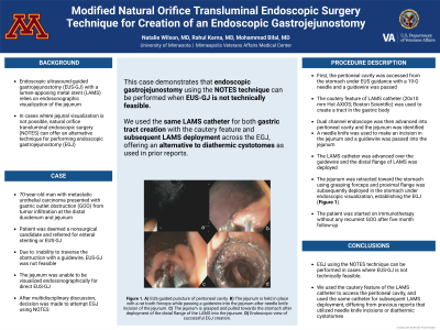
P2846 - Modified Natural Orifice Transluminal Endoscopic Surgery (NOTES) Technique for Creation of an Endoscopic Gastrojejunostomy
Monday, October 28, 2024
10:30 AM - 4:00 PM ET
Location: Exhibit Hall E


Natalie Wilson, MD
University of Minnesota
Minneapolis, MN
Presenting Author(s)
Natalie Wilson, MD1, Rahul Karna, MD2, Mohammad Bilal, MD3
1University of Minnesota, Minneapolis, MN; 2University of Minnesota Medical Center, Minneapolis, MN; 3University of Minnesota and Minneapolis VA Health Care System, Minneapolis, MN
Introduction: Endoscopic ultrasound-guided gastrojejunostomy (EUS-GJ) using a lumen apposing metal stent (LAMS) is an effective treatment for managing gastric outlet obstruction (GOO). EUS-GJ technique depends on endosonographic visualization of the jejunum from either with insufflation of the jejunum with fluid through a orojejunal catheter (OJC) or direct visualization under EUS guidance. In cases where endosonographic visualization of the jejunum is not possible, natural orifice transluminal endoscopic surgery (NOTES) can be used for creation of an endoscopic gastrojejunostomy (EGJ).
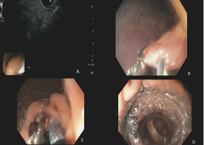
Case Description/Methods: A 70 year old male with history of locally advanced urothelial carcinoma (UCA) of the urinary bladder with previous definitive chemoradiation presented with GOO from metastatic urothelial carcinoma located involving the distal duodenum. Biopsies were consistent with metastatic UCA. Patient was referred for enteral stenting or EUS-GJ. EGD was performed, however, the endoscope or guidewire was unable to traverse the stenosis, and therefore an OJC to distend the jejunum for better visualization of jejunum under EUS guidance was unable to be placed. The procedure was aborted. After multidisciplinary discussion, the patient was deemed to be high risk for surgery and decision was made to proceed with EGJ using NOTES. The serosa was punctured under EUS guidance and a guidewire was placed into the peritoneal cavity. We used the cautery feature of the LAMS to puncture the gastric wall over the guidewire to access the peritoneal cavity. The jejunum was identified, and using a needle knife, an incision was made in the jejunum and a guidewire was passed. Using a dual channel endoscope, a grasping forceps was used to grasp the jejunum in place, and the previously used LAMS catheter was advanced into the jejunum under endoscopic guidance and the distal flange of LAMS was deployed into the jejunum. The grasping forceps was used to pull the jejunum towards the stomach and the proximal flange of the LAMS was deployed in the stomach creating a successful EGJ [Figure 1]. Intraprocedural pneumoperitoneum was managed with intermittent suction. Clear liquid diet was initiated on post operative day 1 and no adverse events occurred.
Discussion: Our case shows that when EUS-GJ is not technically feasible, NOTES can be effectively used in creating an EGJ. We used the cautery feature of the LAMS catheter to access the peritoneal cavity, and used the same catheter for subsequent LAMS deployment.

Disclosures:
Natalie Wilson, MD1, Rahul Karna, MD2, Mohammad Bilal, MD3. P2846 - Modified Natural Orifice Transluminal Endoscopic Surgery (NOTES) Technique for Creation of an Endoscopic Gastrojejunostomy, ACG 2024 Annual Scientific Meeting Abstracts. Philadelphia, PA: American College of Gastroenterology.
1University of Minnesota, Minneapolis, MN; 2University of Minnesota Medical Center, Minneapolis, MN; 3University of Minnesota and Minneapolis VA Health Care System, Minneapolis, MN
Introduction: Endoscopic ultrasound-guided gastrojejunostomy (EUS-GJ) using a lumen apposing metal stent (LAMS) is an effective treatment for managing gastric outlet obstruction (GOO). EUS-GJ technique depends on endosonographic visualization of the jejunum from either with insufflation of the jejunum with fluid through a orojejunal catheter (OJC) or direct visualization under EUS guidance. In cases where endosonographic visualization of the jejunum is not possible, natural orifice transluminal endoscopic surgery (NOTES) can be used for creation of an endoscopic gastrojejunostomy (EGJ).
Case Description/Methods: A 70 year old male with history of locally advanced urothelial carcinoma (UCA) of the urinary bladder with previous definitive chemoradiation presented with GOO from metastatic urothelial carcinoma located involving the distal duodenum. Biopsies were consistent with metastatic UCA. Patient was referred for enteral stenting or EUS-GJ. EGD was performed, however, the endoscope or guidewire was unable to traverse the stenosis, and therefore an OJC to distend the jejunum for better visualization of jejunum under EUS guidance was unable to be placed. The procedure was aborted. After multidisciplinary discussion, the patient was deemed to be high risk for surgery and decision was made to proceed with EGJ using NOTES. The serosa was punctured under EUS guidance and a guidewire was placed into the peritoneal cavity. We used the cautery feature of the LAMS to puncture the gastric wall over the guidewire to access the peritoneal cavity. The jejunum was identified, and using a needle knife, an incision was made in the jejunum and a guidewire was passed. Using a dual channel endoscope, a grasping forceps was used to grasp the jejunum in place, and the previously used LAMS catheter was advanced into the jejunum under endoscopic guidance and the distal flange of LAMS was deployed into the jejunum. The grasping forceps was used to pull the jejunum towards the stomach and the proximal flange of the LAMS was deployed in the stomach creating a successful EGJ [Figure 1]. Intraprocedural pneumoperitoneum was managed with intermittent suction. Clear liquid diet was initiated on post operative day 1 and no adverse events occurred.
Discussion: Our case shows that when EUS-GJ is not technically feasible, NOTES can be effectively used in creating an EGJ. We used the cautery feature of the LAMS catheter to access the peritoneal cavity, and used the same catheter for subsequent LAMS deployment.

Figure: Endoscopic ultrasound guided puncture of the peritoneal cavity (A). Dual channel endoscope being used to hold the jejunum in place with a rat tooth forceps while passing a guidewire into the jejunum after needle knife incision of the jejunum (B). The jejunum is grasped and pulled towards the stomach after deployment of the distal flange of the lumen apposing metal stent into the jejunum (C). Endoscopic view of successful endoscopic gastrojejunostomy creation (D).
Disclosures:
Natalie Wilson indicated no relevant financial relationships.
Rahul Karna indicated no relevant financial relationships.
Mohammad Bilal: Boston Scientific – Consultant. Cook endoscopy – Speakers Bureau.
Natalie Wilson, MD1, Rahul Karna, MD2, Mohammad Bilal, MD3. P2846 - Modified Natural Orifice Transluminal Endoscopic Surgery (NOTES) Technique for Creation of an Endoscopic Gastrojejunostomy, ACG 2024 Annual Scientific Meeting Abstracts. Philadelphia, PA: American College of Gastroenterology.
