Monday Poster Session
Category: Interventional Endoscopy
P2849 - EUS-Guided FNA Diagnosis of Rosai-Dorfman Disease
Monday, October 28, 2024
10:30 AM - 4:00 PM ET
Location: Exhibit Hall E

Has Audio

Talwinder Nagi, MD
Florida International University
Boca Raton, FL
Presenting Author(s)
Talwinder Nagi, MD1, Polina Gaisinskaya, MD2, Peter J. Bross, MD2, Adib Chaaya, MD2
1Florida International University, Boca Raton, FL; 2Cooper Health Gastroenterology, Camden, NJ
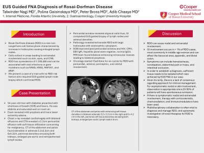
Introduction: Rosai-Dorfman disease (RDD) is a rare non-Langerhans cell histiocytosis characterized by increase in histiocytes causing enlarged lymph nodes. The cells can collect in areas leading to extranodal involvement such as skin, eyes, and CNS. RDD has a prevalence of 1:200,000 and can be associated with viral infections or gene mutations such as NRAS, KRAS, MAP2K1, and ARAF. We present a case of a man with no RDD risk factors who required EUS-guided lymph node biopsy which confirmed RDD.
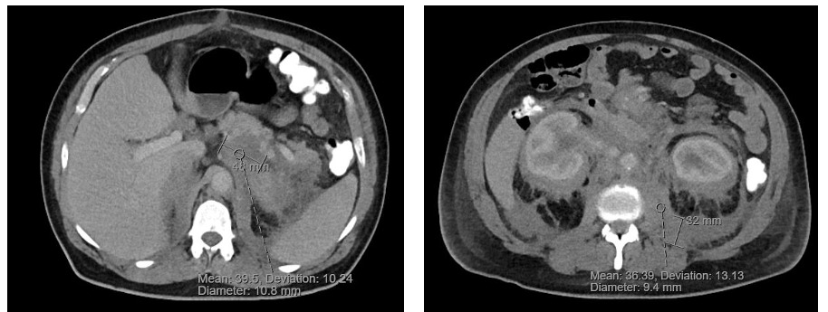
Case Description/Methods: 54-year-old man with diabetes presented with shortness of breath (SOB) and fevers. He was afebrile and saturated well on room air. Exam was pertinent for mild proptosis and trace lower extremity edema. Chest x-ray revealed cardiomegaly with bilateral effusions and CTA revealed a 3.3cm pericardial effusion with soft tissue infiltration concerning for malignancy. CT of the abdomen and pelvis found densities in adrenals 5.2x3.2cm and 6x4.2cm, perirenal densities encasing both kidneys, enlarged pre-aortic and retroperitoneal lymph nodes. Pericardial window revealed atypical cells thus, GI completed EUS guided biopsy of lymph nodes and adrenal densities. Pathology revealed extranodal RDD with large histiocytes with eosinophilic cytoplasm. SOB improved post pericardial window and HIV, CMV, EBV IgM, hepatitis panel were negative, normal IgG4. MRI brain found bilateral enhancing retroocular masses leading to bilateral proptosis. Oncology started Cladribine for six cycles for RDD with pericardial, adrenal, perinephric, and orbital involvement.
Discussion: RDD can cause nodal and extranodal involvement. GI involvement occurs in < 1% of RDD cases, most commonly in middle-age women and can affect the ileocecal area, appendix, and distal colon. Symptoms can include hematochezia, constipation, abdominal pain or mass, and intestinal occlusion. In order to establish a diagnosis, sufficient tissue needs to be isolated which was achieved by EUS FNA in our case. Given its rarity, there is a lack of consensus regarding approach for clinical management. For asymptomatic nodal or skin involvement, observation is appropriate since 20-50% of patients will have spontaneous remission. However if there is symptomatic nodal and extranodal involvement, therapy with corticosteroids, chemoradiation, and immunomodulators have been used. Multidisciplinary collaboration is often vital to diagnose and manage RDD, and systematic investigation of novel therapies for RDD is necessary.

Disclosures:
Talwinder Nagi, MD1, Polina Gaisinskaya, MD2, Peter J. Bross, MD2, Adib Chaaya, MD2. P2849 - EUS-Guided FNA Diagnosis of Rosai-Dorfman Disease, ACG 2024 Annual Scientific Meeting Abstracts. Philadelphia, PA: American College of Gastroenterology.
1Florida International University, Boca Raton, FL; 2Cooper Health Gastroenterology, Camden, NJ
Introduction: Rosai-Dorfman disease (RDD) is a rare non-Langerhans cell histiocytosis characterized by increase in histiocytes causing enlarged lymph nodes. The cells can collect in areas leading to extranodal involvement such as skin, eyes, and CNS. RDD has a prevalence of 1:200,000 and can be associated with viral infections or gene mutations such as NRAS, KRAS, MAP2K1, and ARAF. We present a case of a man with no RDD risk factors who required EUS-guided lymph node biopsy which confirmed RDD.
Case Description/Methods: 54-year-old man with diabetes presented with shortness of breath (SOB) and fevers. He was afebrile and saturated well on room air. Exam was pertinent for mild proptosis and trace lower extremity edema. Chest x-ray revealed cardiomegaly with bilateral effusions and CTA revealed a 3.3cm pericardial effusion with soft tissue infiltration concerning for malignancy. CT of the abdomen and pelvis found densities in adrenals 5.2x3.2cm and 6x4.2cm, perirenal densities encasing both kidneys, enlarged pre-aortic and retroperitoneal lymph nodes. Pericardial window revealed atypical cells thus, GI completed EUS guided biopsy of lymph nodes and adrenal densities. Pathology revealed extranodal RDD with large histiocytes with eosinophilic cytoplasm. SOB improved post pericardial window and HIV, CMV, EBV IgM, hepatitis panel were negative, normal IgG4. MRI brain found bilateral enhancing retroocular masses leading to bilateral proptosis. Oncology started Cladribine for six cycles for RDD with pericardial, adrenal, perinephric, and orbital involvement.
Discussion: RDD can cause nodal and extranodal involvement. GI involvement occurs in < 1% of RDD cases, most commonly in middle-age women and can affect the ileocecal area, appendix, and distal colon. Symptoms can include hematochezia, constipation, abdominal pain or mass, and intestinal occlusion. In order to establish a diagnosis, sufficient tissue needs to be isolated which was achieved by EUS FNA in our case. Given its rarity, there is a lack of consensus regarding approach for clinical management. For asymptomatic nodal or skin involvement, observation is appropriate since 20-50% of patients will have spontaneous remission. However if there is symptomatic nodal and extranodal involvement, therapy with corticosteroids, chemoradiation, and immunomodulators have been used. Multidisciplinary collaboration is often vital to diagnose and manage RDD, and systematic investigation of novel therapies for RDD is necessary.

Figure: CT of the abdomen and pelvis with enhancing soft tissue densities in bilateral adrenals 5.2 x 3.2 cm in the right and 6 x 4.2 cm in the left, perirenal soft tissue densities encasing both kidneys, enlarged pre-aortic lymph node 4.8 cm.
Disclosures:
Talwinder Nagi indicated no relevant financial relationships.
Polina Gaisinskaya indicated no relevant financial relationships.
Peter Bross indicated no relevant financial relationships.
Adib Chaaya indicated no relevant financial relationships.
Talwinder Nagi, MD1, Polina Gaisinskaya, MD2, Peter J. Bross, MD2, Adib Chaaya, MD2. P2849 - EUS-Guided FNA Diagnosis of Rosai-Dorfman Disease, ACG 2024 Annual Scientific Meeting Abstracts. Philadelphia, PA: American College of Gastroenterology.
