Tuesday Poster Session
Category: IBD
P4446 - Differentiating Mesenteric Panniculitis From Crohn’s Disease
Tuesday, October 29, 2024
10:30 AM - 4:00 PM ET
Location: Exhibit Hall E

Has Audio

Mansi Sheth, DO
Jefferson Torresdale Hospital
Bridgewater, NJ
Presenting Author(s)
Mansi Sheth, DO, Salman Qureshi, DO, Chad Gunsolly, DO
Jefferson Torresdale Hospital, Philadelphia, PA
Introduction: Mesenteric panniculitis (MP), a condition of chronic inflammation leading to mesenteric fat necrosis and/or fibrosis, has been studied as a disease of various etiologies without a clear cause. Several articles have proposed various immune-related pathologies as risk factors of MP, however acute pathologies such as abdominal surgery, trauma, and malignancy have been implicated as well. Undiagnosed or untreated MP can be fatal. Crohn’s Disease (CD) is an immunologic inflammatory condition that chronically impacts the gastrointestinal tract. In the initial stages, CD has a similar presentation to MP including abdominal pain, bloody diarrhea, nausea, and vomiting. Colonoscopy and tissue pathology evaluation is vital for proper diagnosis and continued treatment.
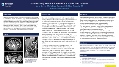
Case Description/Methods: Our patient is a 20-year-old male with no past medical history who presented twice in 2 days for abdominal pain, worsening diarrhea, and 2 episodes of hematochezia. He had two prior episodes of crampy abdominal pain and bloody diarrhea with previous imaging demonstrating terminal ileal thickening and labs significant for elevated C-reactive protein. A colonoscopy was never performed previously. During this visit, he was febrile, tachycardic, and tachypneic with diffuse abdominal pain, nausea, vomiting, and hematochezia. Abdominal CT scan showed multiple central mesenteric lymph nodes with surrounding inflammatory changes thought to be mesenteric panniculitis, as well as hepatosplenomegaly and no evidence of colitis. He was admitted for sepsis of unknown source and empirically treated with antibiotics. Colonoscopy demonstrated normal mucosa of the entire bowel and nodularity of the terminal ilium without prominent ulcerations. Pathology showed microscopic noncaseating granuloma and follicular hyperplasia of the terminal ileum indicative of Crohn’s disease. Additional studies for celiac, inflammatory, infectious, and autoimmune etiologies were negative.
Discussion: Mesenteric Panniculitis and Crohn’s Disease may appear with similar presentations such as severe abdominal pain, nausea, and vomiting. Initial workup of imaging (CT) and laboratory work can differentiate the two conditions. Further studies like surgical biopsy and tissue evaluation for MP and colonoscopy for CD are standard diagnostic modalities. While both MP and CD can be treated with anti-inflammatory and immunosuppressive agents, the importance of proper diagnosis is crucial for long-term treatment.
Disclosures:
Mansi Sheth, DO, Salman Qureshi, DO, Chad Gunsolly, DO. P4446 - Differentiating Mesenteric Panniculitis From Crohn’s Disease, ACG 2024 Annual Scientific Meeting Abstracts. Philadelphia, PA: American College of Gastroenterology.
Jefferson Torresdale Hospital, Philadelphia, PA
Introduction: Mesenteric panniculitis (MP), a condition of chronic inflammation leading to mesenteric fat necrosis and/or fibrosis, has been studied as a disease of various etiologies without a clear cause. Several articles have proposed various immune-related pathologies as risk factors of MP, however acute pathologies such as abdominal surgery, trauma, and malignancy have been implicated as well. Undiagnosed or untreated MP can be fatal. Crohn’s Disease (CD) is an immunologic inflammatory condition that chronically impacts the gastrointestinal tract. In the initial stages, CD has a similar presentation to MP including abdominal pain, bloody diarrhea, nausea, and vomiting. Colonoscopy and tissue pathology evaluation is vital for proper diagnosis and continued treatment.
Case Description/Methods: Our patient is a 20-year-old male with no past medical history who presented twice in 2 days for abdominal pain, worsening diarrhea, and 2 episodes of hematochezia. He had two prior episodes of crampy abdominal pain and bloody diarrhea with previous imaging demonstrating terminal ileal thickening and labs significant for elevated C-reactive protein. A colonoscopy was never performed previously. During this visit, he was febrile, tachycardic, and tachypneic with diffuse abdominal pain, nausea, vomiting, and hematochezia. Abdominal CT scan showed multiple central mesenteric lymph nodes with surrounding inflammatory changes thought to be mesenteric panniculitis, as well as hepatosplenomegaly and no evidence of colitis. He was admitted for sepsis of unknown source and empirically treated with antibiotics. Colonoscopy demonstrated normal mucosa of the entire bowel and nodularity of the terminal ilium without prominent ulcerations. Pathology showed microscopic noncaseating granuloma and follicular hyperplasia of the terminal ileum indicative of Crohn’s disease. Additional studies for celiac, inflammatory, infectious, and autoimmune etiologies were negative.
Discussion: Mesenteric Panniculitis and Crohn’s Disease may appear with similar presentations such as severe abdominal pain, nausea, and vomiting. Initial workup of imaging (CT) and laboratory work can differentiate the two conditions. Further studies like surgical biopsy and tissue evaluation for MP and colonoscopy for CD are standard diagnostic modalities. While both MP and CD can be treated with anti-inflammatory and immunosuppressive agents, the importance of proper diagnosis is crucial for long-term treatment.
Disclosures:
Mansi Sheth indicated no relevant financial relationships.
Salman Qureshi indicated no relevant financial relationships.
Chad Gunsolly indicated no relevant financial relationships.
Mansi Sheth, DO, Salman Qureshi, DO, Chad Gunsolly, DO. P4446 - Differentiating Mesenteric Panniculitis From Crohn’s Disease, ACG 2024 Annual Scientific Meeting Abstracts. Philadelphia, PA: American College of Gastroenterology.
