Tuesday Poster Session
Category: Interventional Endoscopy
P4525 - Unraveling an Uncommon Cause of Hemobilia: An Interesting Case of Gallbladder Adenocarcinoma
Tuesday, October 29, 2024
10:30 AM - 4:00 PM ET
Location: Exhibit Hall E

Has Audio
- SP
Spyros Peppas, MD
MedStar Health-Georgetown/Washington Hospital Center
Washington, DC
Presenting Author(s)
Spyridon Peppas, MD1, Srikar Reddy, MD2, Sitharthan Sekar, MD2, Nadim G. Haddad, MD2
1MedStar Health-Georgetown/Washington Hospital Center, Washington, DC; 2MedStar Georgetown University Hospital, Washington, DC
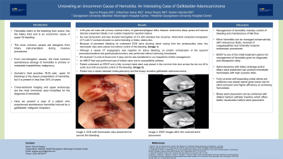
Introduction: Hemobilia is a rare presentation of gallbladder cancer. The majority of hemobilia cases are iatrogenic caused by invasive pancreatobiliary procedures with all-cause malignancies accounting for 10% of total cases. Here we present a case of a patient who experienced spontaneous hemobilia induced by a gallbladder malignant neoplasm.
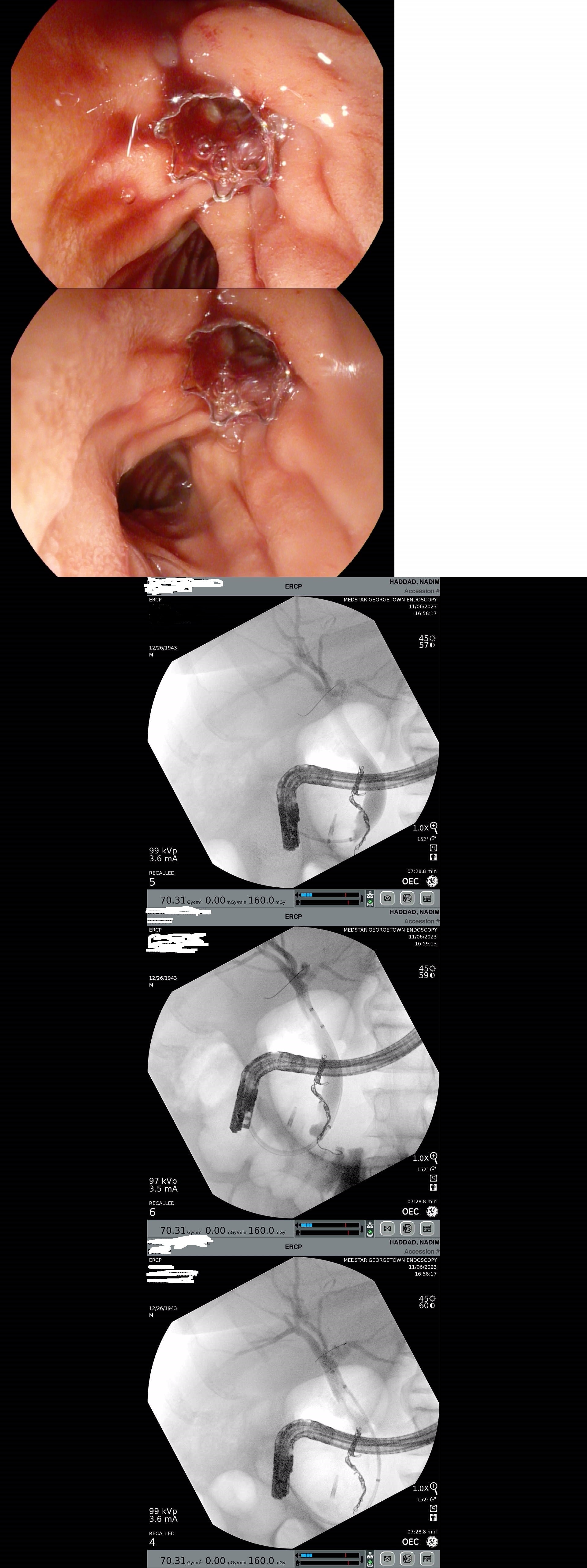
Case Description/Methods: A 80-year-old male with primary medical history of gastroesophageal reflux disease, obstructive sleep apnea and seizure disorder presented initially in an outside hospital for reported melena, abdominal pain and fatigue. At presentation, he was tachycardic and labs showed hemoglobin of 4.0 with elevated liver enzymes. Abdominal computed tomography (CT) with IV contrast showed no active bleeding or biliary obstruction. He underwent an EGD showing blood oozing from the periampullary area and two clips were placed for control of the bleeding. Given persistent bleeding, he had a repeat EGD which showed appropriate position of the clips, fresh blood in the duodenal lumen, but it was unclear where the origin of the bleeding was and was presumed to be related to a bleeding ulcer. Although a repeat CT angiography was negative for active bleeding, an empiric embolization of the superior pancreatoduodenal and gastroduodenal artery was performed without achieving hemostasis. He received 12 units of blood over 5 days and he was transferred to our hospital for further management. An MRCP that was performed was of limited value due to susceptibility artifacts. In the following day, patient underwent an ERCP which revealed fresh blood with clots flowing from the gallbladder to the ampulla of Vater. A fully covered metal stent was placed in the common bile duct across the low cut off to cystic duct with successful control of the bleeding. Subsequently, patient had a robotic-assisted cholecystectomy and the biopsy revealed gallbladder adenocarcinoma.
Discussion: Gallbladder neoplasms are difficult to diagnose and distinguish. Although in the majority of cases they present with non-specific and subtle symptoms, rarely may manifest with biliary obstruction and hemobilia. In this case, gallbladder neoplasm was not evident in multiple cross-sectional imaging studies and ERCP revealed the gallbladder as the original site of bleeding with a successful placement of a metal stent at the cystic duct orifice and achievement of hemostasis.

Disclosures:
Spyridon Peppas, MD1, Srikar Reddy, MD2, Sitharthan Sekar, MD2, Nadim G. Haddad, MD2. P4525 - Unraveling an Uncommon Cause of Hemobilia: An Interesting Case of Gallbladder Adenocarcinoma, ACG 2024 Annual Scientific Meeting Abstracts. Philadelphia, PA: American College of Gastroenterology.
1MedStar Health-Georgetown/Washington Hospital Center, Washington, DC; 2MedStar Georgetown University Hospital, Washington, DC
Introduction: Hemobilia is a rare presentation of gallbladder cancer. The majority of hemobilia cases are iatrogenic caused by invasive pancreatobiliary procedures with all-cause malignancies accounting for 10% of total cases. Here we present a case of a patient who experienced spontaneous hemobilia induced by a gallbladder malignant neoplasm.
Case Description/Methods: A 80-year-old male with primary medical history of gastroesophageal reflux disease, obstructive sleep apnea and seizure disorder presented initially in an outside hospital for reported melena, abdominal pain and fatigue. At presentation, he was tachycardic and labs showed hemoglobin of 4.0 with elevated liver enzymes. Abdominal computed tomography (CT) with IV contrast showed no active bleeding or biliary obstruction. He underwent an EGD showing blood oozing from the periampullary area and two clips were placed for control of the bleeding. Given persistent bleeding, he had a repeat EGD which showed appropriate position of the clips, fresh blood in the duodenal lumen, but it was unclear where the origin of the bleeding was and was presumed to be related to a bleeding ulcer. Although a repeat CT angiography was negative for active bleeding, an empiric embolization of the superior pancreatoduodenal and gastroduodenal artery was performed without achieving hemostasis. He received 12 units of blood over 5 days and he was transferred to our hospital for further management. An MRCP that was performed was of limited value due to susceptibility artifacts. In the following day, patient underwent an ERCP which revealed fresh blood with clots flowing from the gallbladder to the ampulla of Vater. A fully covered metal stent was placed in the common bile duct across the low cut off to cystic duct with successful control of the bleeding. Subsequently, patient had a robotic-assisted cholecystectomy and the biopsy revealed gallbladder adenocarcinoma.
Discussion: Gallbladder neoplasms are difficult to diagnose and distinguish. Although in the majority of cases they present with non-specific and subtle symptoms, rarely may manifest with biliary obstruction and hemobilia. In this case, gallbladder neoplasm was not evident in multiple cross-sectional imaging studies and ERCP revealed the gallbladder as the original site of bleeding with a successful placement of a metal stent at the cystic duct orifice and achievement of hemostasis.

Figure: ERCP images with covered stent placement
Disclosures:
Spyridon Peppas indicated no relevant financial relationships.
Srikar Reddy indicated no relevant financial relationships.
Sitharthan Sekar indicated no relevant financial relationships.
Nadim Haddad indicated no relevant financial relationships.
Spyridon Peppas, MD1, Srikar Reddy, MD2, Sitharthan Sekar, MD2, Nadim G. Haddad, MD2. P4525 - Unraveling an Uncommon Cause of Hemobilia: An Interesting Case of Gallbladder Adenocarcinoma, ACG 2024 Annual Scientific Meeting Abstracts. Philadelphia, PA: American College of Gastroenterology.
