Tuesday Poster Session
Category: Interventional Endoscopy
P4528 - Diagnostic Utility of Endoscopic Ultrasound in Identifying a Rare Celiac Axis Mass: Organizing Hematoma From Spontaneous Contained Rupture
Tuesday, October 29, 2024
10:30 AM - 4:00 PM ET
Location: Exhibit Hall E

Has Audio

Harneet Sangha, BS
Washington State University Elson S. Floyd College of Medicine
Spokane, WA
Presenting Author(s)
Harneet Sangha, BS1, Tavishi Girotra, MBBS2, Shreya Chopra, MBBS2, Navansh Johnson, 2, Anusha Singhania, MBBS2, Mohit Girotra, MD1
1Washington State University Elson S. Floyd College of Medicine, Spokane, WA; 2Digestive Health Institute, Swedish Medical Center, Seattle, WA
Introduction: Celiac axis masses are rare and pose considerable diagnostic and therapeutic challenges due to their intricate anatomical location. Accurate diagnosis is imperative, as these masses may represent benign conditions such as retroperitoneal fibrosis, but they can also be malignant or premalignant processes.
We present a case to highlight the utility of Endoscopic Ultrasound (EUS) in diagnosis of a newly emerged and symptomatic celiac mass.
Case Description/Methods: A 53-year-old man with previous uncomplicated diverticulosis and adenomatous polyps, presented to the ED with a month-long history of constant mid-epigastric pain, not associated with fever, unintentional loss of weight/appetite, jaundice, nausea, or diarrhea. Computerized Tomography (CT) revealed an abnormal irregular soft tissue mass in the upper retroperitoneum, in close association with celiac trunk, and was referred for EUS. Previous CT from a year ago did not reveal any similar abnormality.
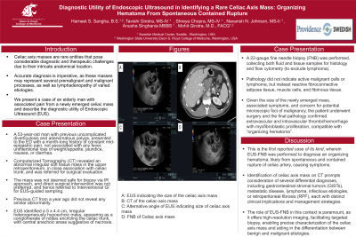
EUS identified a 5x4.4 cm, irregular heterogeneously hypoechoic/anechoic mass, appearing as a conglomerate of nodes encircling the celiac trunk, with central anechoic areas suggestive of necrosis. A 22-gauge fine needle biopsy (FNB) was performed, collecting both fluid and tissue samples for histology and flow cytometry. The biopsies did not indicate active malignant cells or lymphoma, but instead reactive fibroconnective adipose tissue, muscle cells, and fibrinous tissue.
Given the size of the newly emerged mass, associated symptoms, and concern for potential microscopic foci of malignancy, the patient underwent surgery and final pathology confirmed extravascular and intravascular thrombi/hemorrhage with myofibroblastic proliferation, compatible with organizing hematoma.
Discussion: This is the first reported case of its kind, wherein EUS-FNB was performed to diagnose an organizing hematoma, likely from spontaneous and contained rupture of celiac artery, causing symptoms.
Identification of celiac axis mass on CT prompts consideration of several differential diagnoses, including gastrointestinal stromal tumors (GISTs), metastatic disease, lymphoma, infectious etiologies, or retroperitoneal fibrosis (RPF), each with distinct clinical implications and management strategies. The role of EUS in this context is paramount, as it offers high-resolution imaging, facilitating targeted biopsy, enabling precise characterization of the celiac axis mass and aiding in the differentiation between benign and malignant etiologies.

Disclosures:
Harneet Sangha, BS1, Tavishi Girotra, MBBS2, Shreya Chopra, MBBS2, Navansh Johnson, 2, Anusha Singhania, MBBS2, Mohit Girotra, MD1. P4528 - Diagnostic Utility of Endoscopic Ultrasound in Identifying a Rare Celiac Axis Mass: Organizing Hematoma From Spontaneous Contained Rupture, ACG 2024 Annual Scientific Meeting Abstracts. Philadelphia, PA: American College of Gastroenterology.
1Washington State University Elson S. Floyd College of Medicine, Spokane, WA; 2Digestive Health Institute, Swedish Medical Center, Seattle, WA
Introduction: Celiac axis masses are rare and pose considerable diagnostic and therapeutic challenges due to their intricate anatomical location. Accurate diagnosis is imperative, as these masses may represent benign conditions such as retroperitoneal fibrosis, but they can also be malignant or premalignant processes.
We present a case to highlight the utility of Endoscopic Ultrasound (EUS) in diagnosis of a newly emerged and symptomatic celiac mass.
Case Description/Methods: A 53-year-old man with previous uncomplicated diverticulosis and adenomatous polyps, presented to the ED with a month-long history of constant mid-epigastric pain, not associated with fever, unintentional loss of weight/appetite, jaundice, nausea, or diarrhea. Computerized Tomography (CT) revealed an abnormal irregular soft tissue mass in the upper retroperitoneum, in close association with celiac trunk, and was referred for EUS. Previous CT from a year ago did not reveal any similar abnormality.
EUS identified a 5x4.4 cm, irregular heterogeneously hypoechoic/anechoic mass, appearing as a conglomerate of nodes encircling the celiac trunk, with central anechoic areas suggestive of necrosis. A 22-gauge fine needle biopsy (FNB) was performed, collecting both fluid and tissue samples for histology and flow cytometry. The biopsies did not indicate active malignant cells or lymphoma, but instead reactive fibroconnective adipose tissue, muscle cells, and fibrinous tissue.
Given the size of the newly emerged mass, associated symptoms, and concern for potential microscopic foci of malignancy, the patient underwent surgery and final pathology confirmed extravascular and intravascular thrombi/hemorrhage with myofibroblastic proliferation, compatible with organizing hematoma.
Discussion: This is the first reported case of its kind, wherein EUS-FNB was performed to diagnose an organizing hematoma, likely from spontaneous and contained rupture of celiac artery, causing symptoms.
Identification of celiac axis mass on CT prompts consideration of several differential diagnoses, including gastrointestinal stromal tumors (GISTs), metastatic disease, lymphoma, infectious etiologies, or retroperitoneal fibrosis (RPF), each with distinct clinical implications and management strategies. The role of EUS in this context is paramount, as it offers high-resolution imaging, facilitating targeted biopsy, enabling precise characterization of the celiac axis mass and aiding in the differentiation between benign and malignant etiologies.

Figure: A: EUS indicating the size of the celiac axis mass; B: CT of the celiac axis mass; C: Alternative angle of EUS indicating size of celiac axis mass; D: FNB of Celiac axis mass
Disclosures:
Harneet Sangha indicated no relevant financial relationships.
Tavishi Girotra indicated no relevant financial relationships.
Shreya Chopra indicated no relevant financial relationships.
Navansh Johnson indicated no relevant financial relationships.
Anusha Singhania indicated no relevant financial relationships.
Mohit Girotra indicated no relevant financial relationships.
Harneet Sangha, BS1, Tavishi Girotra, MBBS2, Shreya Chopra, MBBS2, Navansh Johnson, 2, Anusha Singhania, MBBS2, Mohit Girotra, MD1. P4528 - Diagnostic Utility of Endoscopic Ultrasound in Identifying a Rare Celiac Axis Mass: Organizing Hematoma From Spontaneous Contained Rupture, ACG 2024 Annual Scientific Meeting Abstracts. Philadelphia, PA: American College of Gastroenterology.
