Tuesday Poster Session
Category: Interventional Endoscopy
P4539 - An Uncommon Result in a Common Finding: The Rare Case of a Gastric Glomus Tumor
Tuesday, October 29, 2024
10:30 AM - 4:00 PM ET
Location: Exhibit Hall E

Has Audio

Samantha A. Menegas, MD
Duke University Hospital
Durham, NC
Presenting Author(s)
Samantha A. Menegas, MD, Pedro A. Rosa-Cortés, MD, Rebecca A. Burbridge, MD
Duke University Hospital, Durham, NC
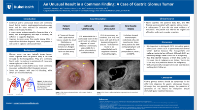
Introduction: Incidental gastric submucosal lesions are commonly found during routine esophagogastroduodenoscopy (EGD) and are often referred for evaluation with endoscopic ultrasound (EUS). In most cases, endosonographic characteristics such as echogenicity and layer of invasion are sufficient to suggest a diagnosis. Occasionally, fine needle biopsy (FNB) is required for a definitive diagnosis. This is the case of a rare result in a gastric submucosal lesion.
Case Description/Methods: A 75 year-old female underwent an EGD due to iron-deficiency anemia. Findings showed a submucosal lesion in the antrum. An EUS showed an isoechoic, 2 cm by 1.3 cm lesion originating in the muscularis propria. FNB was performed. Pathology showed monomorphic epithelioid cells with round nuclei. Stains were positive for SMA and synaptophysin and negative for pancytokeratin, INSM1 and CD45. These findings were consistent with a gastric glomus tumor (GGT). A CT scan showed no metastasis and the patient was referred to surgery, ultimately undergoing a wedge resection given bleeding potential.
Discussion: Glomus tumors are rare, typically benign tumors that originate from the glomus body. They are commonly found under the nails or in peripheral soft tissue and are rarely found in the gastrointestinal (GI) tract. GGTs occur more commonly in women and tend to affect the gastric antrum. Some present with GI bleeding while others are discovered incidentally. GGTs may be seen on EGD as a smooth mucosal protrusion. EUS can be used to demonstrate the tumor’s layer of origin and to obtain tissue for pathologic analysis, which is necessary to make a definitive diagnosis. Immunohistochemical staining will often show positivity for SMA and vimentin and will be negative for markers present in other lesions. It is important to distinguish GGTs from other gastric submucosal tumors such as gastrointestinal stromal tumors (GISTs) because the management and malignant potential are different. Although GGTs are commonly benign, there is still a risk of malignancy. Data that identify features of GGTs that confer increased risk of malignancy are limited. Tumor size ≥5 cm may be a predictive feature for malignancy. GGTs are generally managed with wide local excision and long-term follow up. GGTs should always be considered in the differential diagnosis of gastric submucosal lesions. Although mostly benign, the presence of symptoms or risk factors for malignancy should guide decision-making for definitive treatment.

Disclosures:
Samantha A. Menegas, MD, Pedro A. Rosa-Cortés, MD, Rebecca A. Burbridge, MD. P4539 - An Uncommon Result in a Common Finding: The Rare Case of a Gastric Glomus Tumor, ACG 2024 Annual Scientific Meeting Abstracts. Philadelphia, PA: American College of Gastroenterology.
Duke University Hospital, Durham, NC
Introduction: Incidental gastric submucosal lesions are commonly found during routine esophagogastroduodenoscopy (EGD) and are often referred for evaluation with endoscopic ultrasound (EUS). In most cases, endosonographic characteristics such as echogenicity and layer of invasion are sufficient to suggest a diagnosis. Occasionally, fine needle biopsy (FNB) is required for a definitive diagnosis. This is the case of a rare result in a gastric submucosal lesion.
Case Description/Methods: A 75 year-old female underwent an EGD due to iron-deficiency anemia. Findings showed a submucosal lesion in the antrum. An EUS showed an isoechoic, 2 cm by 1.3 cm lesion originating in the muscularis propria. FNB was performed. Pathology showed monomorphic epithelioid cells with round nuclei. Stains were positive for SMA and synaptophysin and negative for pancytokeratin, INSM1 and CD45. These findings were consistent with a gastric glomus tumor (GGT). A CT scan showed no metastasis and the patient was referred to surgery, ultimately undergoing a wedge resection given bleeding potential.
Discussion: Glomus tumors are rare, typically benign tumors that originate from the glomus body. They are commonly found under the nails or in peripheral soft tissue and are rarely found in the gastrointestinal (GI) tract. GGTs occur more commonly in women and tend to affect the gastric antrum. Some present with GI bleeding while others are discovered incidentally. GGTs may be seen on EGD as a smooth mucosal protrusion. EUS can be used to demonstrate the tumor’s layer of origin and to obtain tissue for pathologic analysis, which is necessary to make a definitive diagnosis. Immunohistochemical staining will often show positivity for SMA and vimentin and will be negative for markers present in other lesions. It is important to distinguish GGTs from other gastric submucosal tumors such as gastrointestinal stromal tumors (GISTs) because the management and malignant potential are different. Although GGTs are commonly benign, there is still a risk of malignancy. Data that identify features of GGTs that confer increased risk of malignancy are limited. Tumor size ≥5 cm may be a predictive feature for malignancy. GGTs are generally managed with wide local excision and long-term follow up. GGTs should always be considered in the differential diagnosis of gastric submucosal lesions. Although mostly benign, the presence of symptoms or risk factors for malignancy should guide decision-making for definitive treatment.

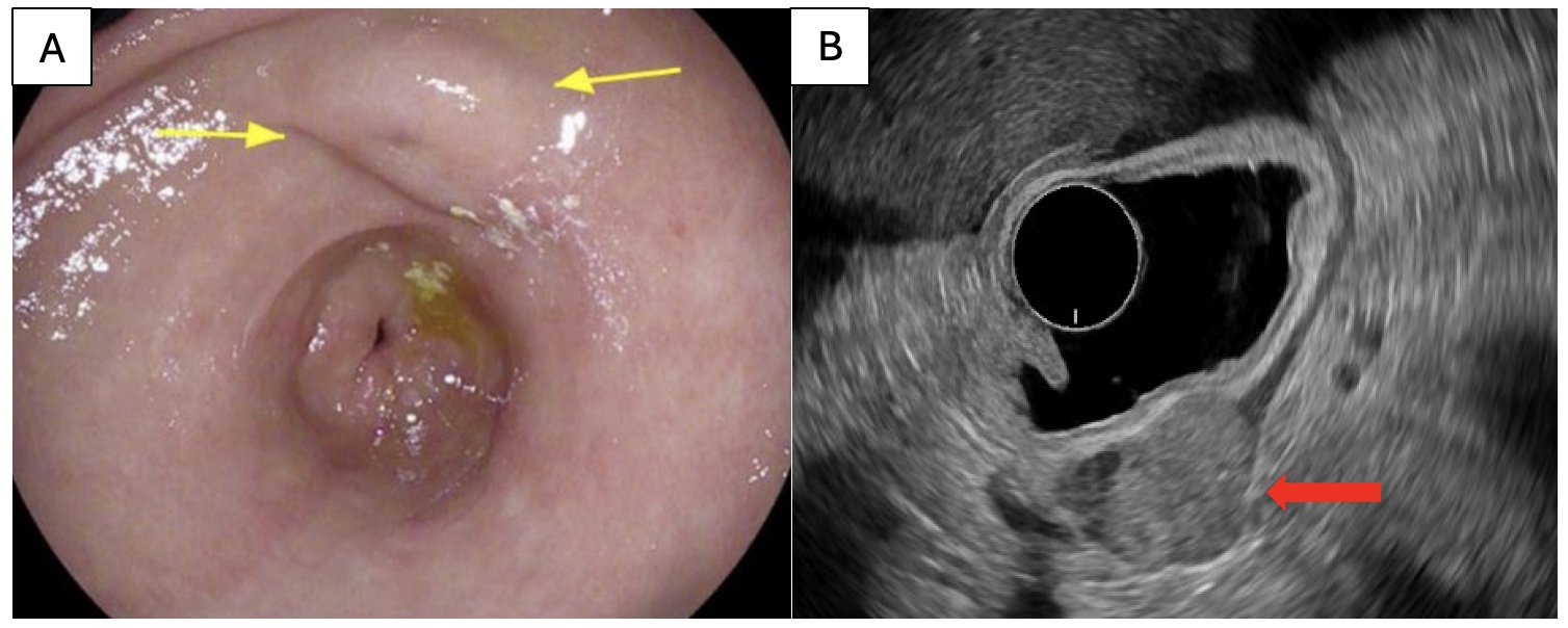
Figure: A. EGD finding of a medium-sized, submucosal, non-circumferential mass with a dimple seen in the gastric antrum
B. EUS finding of an oval intramural lesion, appearing to originate from the muscularis propria
B. EUS finding of an oval intramural lesion, appearing to originate from the muscularis propria
Disclosures:
Samantha Menegas indicated no relevant financial relationships.
Pedro Rosa-Cortés indicated no relevant financial relationships.
Rebecca Burbridge indicated no relevant financial relationships.
Samantha A. Menegas, MD, Pedro A. Rosa-Cortés, MD, Rebecca A. Burbridge, MD. P4539 - An Uncommon Result in a Common Finding: The Rare Case of a Gastric Glomus Tumor, ACG 2024 Annual Scientific Meeting Abstracts. Philadelphia, PA: American College of Gastroenterology.
