Tuesday Poster Session
Category: Interventional Endoscopy
P4543 - Treatment of Refractory Duodenal Perforation With Over-the-Scope Stent
Tuesday, October 29, 2024
10:30 AM - 4:00 PM ET
Location: Exhibit Hall E

Has Audio

Omar T. Ahmed, MD
University of Illinois at Chicago
Chicago, IL
Presenting Author(s)
Omar T. Ahmed, MD1, Dexter Nwachukwu, MD1, Dirin Ukwade, MD2, Brian R. Boulay, MD, MPH3
1University of Illinois at Chicago, Chicago, IL; 2University of Illinois College of Medicine, Chicago, IL; 3University of Illinois, Chicago, IL
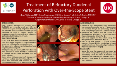
Introduction: Over-the-wire fully covered self-expanding metallic stent (fcSEMS) delivery has been used in treatment of upper GI tract perforations and postoperative leaks. The delivery catheters are generally short and stiff, and therefore, unable to reach distal parts of the GI tract or cross severely angulated strictures. Duodenal stents are only produced as uncovered stents for malignant stricture. In our case, we describe using over-the-scope-stent (OTSS) technique to place a fcSEMS through an angulated area and successfully treat a duodenal perforation that failed surgical repair.
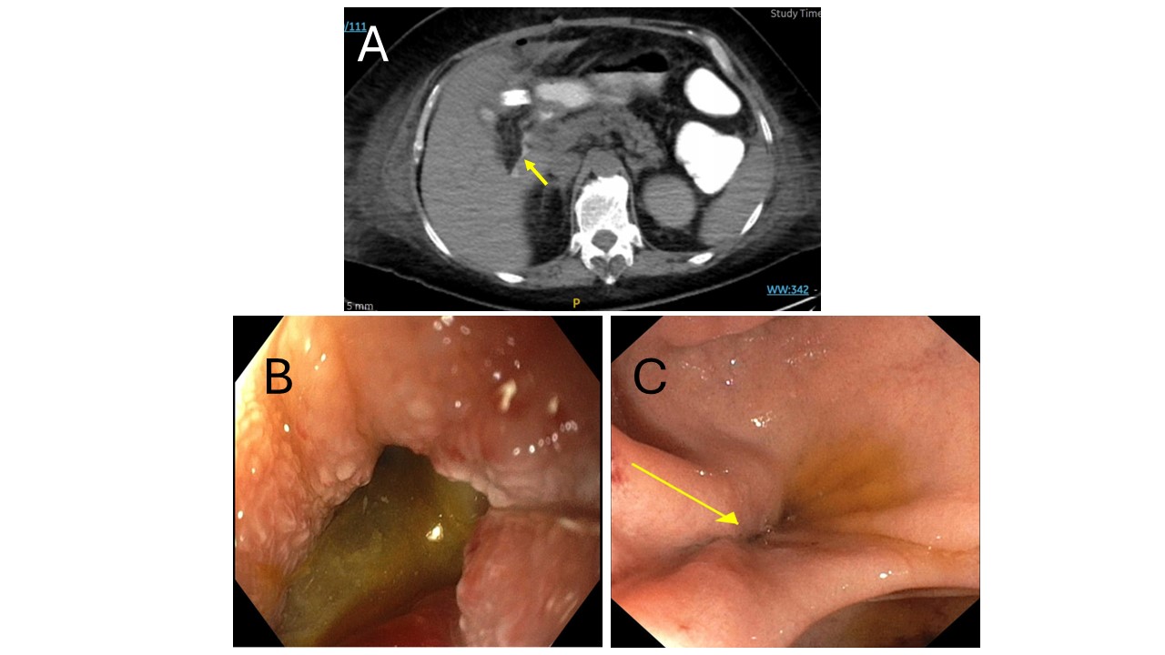
Case Description/Methods: A 40-year-old female with a remote history of ectopic pregnancy was transferred from an outside hospital for partial small bowel obstruction in the setting of a new large ovarian mass. There was evidence of pneumoperitoneum on repeat imaging, and patient was in septic shock and had suffered a cardiac arrest with achievement of ROSC. Exploratory laparotomy revealed a 3cm duodenal ulcer perforation requiring a Graham patch. Three days later, she underwent resection of the ovarian mass. Postoperatively, she was noted to have a continued duodenal leak on upper GI series. Repeat exploratory laparotomy revealed a residual 1cm duodenal perforation. Later, bilious fluid was noted in JP drains and CT confirmed contrast leak from the duodenum(Fig A). EGD was performed and showed a 10mm anterior wall duodenal bulb perforation(Fig B). A 20mm x 10cm fully covered esophageal stent was deployed outside of the patient, and a therapeutic gastroscope was inserted through the stent body where the distal string was grasped with a rat tooth forceps. Withdrawal of the forceps into the working channel allowed us to advance the stent to the duodenum using an over-the-scope stent (OTSS) technique and the stent was successfully positioned. Stent was clipped into place in the antrum using four through-the-scope metal clips. Daily KUBs were obtained to monitor for stent migration. Repeat EGD a month later showed complete healing of the duodenal perforation and the stent was removed(Fig C).
Discussion:
Despite two surgical attempts to patch the duodenal perforation, our patient continued to demonstrate evidence of duodenal leakage. Endoscopic OTSS was eventually successful at stenting over the perforation with placement of anchoring clips to decrease risk of stent migration. Our case provides evidence that OTSS remains a potentially effective, minimally invasive and reliable treatment option for challenging GI tract perforations.

Disclosures:
Omar T. Ahmed, MD1, Dexter Nwachukwu, MD1, Dirin Ukwade, MD2, Brian R. Boulay, MD, MPH3. P4543 - Treatment of Refractory Duodenal Perforation With Over-the-Scope Stent, ACG 2024 Annual Scientific Meeting Abstracts. Philadelphia, PA: American College of Gastroenterology.
1University of Illinois at Chicago, Chicago, IL; 2University of Illinois College of Medicine, Chicago, IL; 3University of Illinois, Chicago, IL
Introduction: Over-the-wire fully covered self-expanding metallic stent (fcSEMS) delivery has been used in treatment of upper GI tract perforations and postoperative leaks. The delivery catheters are generally short and stiff, and therefore, unable to reach distal parts of the GI tract or cross severely angulated strictures. Duodenal stents are only produced as uncovered stents for malignant stricture. In our case, we describe using over-the-scope-stent (OTSS) technique to place a fcSEMS through an angulated area and successfully treat a duodenal perforation that failed surgical repair.
Case Description/Methods: A 40-year-old female with a remote history of ectopic pregnancy was transferred from an outside hospital for partial small bowel obstruction in the setting of a new large ovarian mass. There was evidence of pneumoperitoneum on repeat imaging, and patient was in septic shock and had suffered a cardiac arrest with achievement of ROSC. Exploratory laparotomy revealed a 3cm duodenal ulcer perforation requiring a Graham patch. Three days later, she underwent resection of the ovarian mass. Postoperatively, she was noted to have a continued duodenal leak on upper GI series. Repeat exploratory laparotomy revealed a residual 1cm duodenal perforation. Later, bilious fluid was noted in JP drains and CT confirmed contrast leak from the duodenum(Fig A). EGD was performed and showed a 10mm anterior wall duodenal bulb perforation(Fig B). A 20mm x 10cm fully covered esophageal stent was deployed outside of the patient, and a therapeutic gastroscope was inserted through the stent body where the distal string was grasped with a rat tooth forceps. Withdrawal of the forceps into the working channel allowed us to advance the stent to the duodenum using an over-the-scope stent (OTSS) technique and the stent was successfully positioned. Stent was clipped into place in the antrum using four through-the-scope metal clips. Daily KUBs were obtained to monitor for stent migration. Repeat EGD a month later showed complete healing of the duodenal perforation and the stent was removed(Fig C).
Discussion:
Despite two surgical attempts to patch the duodenal perforation, our patient continued to demonstrate evidence of duodenal leakage. Endoscopic OTSS was eventually successful at stenting over the perforation with placement of anchoring clips to decrease risk of stent migration. Our case provides evidence that OTSS remains a potentially effective, minimally invasive and reliable treatment option for challenging GI tract perforations.

Figure: Fig. A: CT with evidence of contrast leak from the duodenum.
Fig. B: A 10mm anterior wall duodenal bulb perforation seen on EGD.
Fig. C: Healed duodenal perforation on repeat EGD one month later
Fig. B: A 10mm anterior wall duodenal bulb perforation seen on EGD.
Fig. C: Healed duodenal perforation on repeat EGD one month later
Disclosures:
Omar Ahmed indicated no relevant financial relationships.
Dexter Nwachukwu indicated no relevant financial relationships.
Dirin Ukwade indicated no relevant financial relationships.
Brian Boulay indicated no relevant financial relationships.
Omar T. Ahmed, MD1, Dexter Nwachukwu, MD1, Dirin Ukwade, MD2, Brian R. Boulay, MD, MPH3. P4543 - Treatment of Refractory Duodenal Perforation With Over-the-Scope Stent, ACG 2024 Annual Scientific Meeting Abstracts. Philadelphia, PA: American College of Gastroenterology.
