Tuesday Poster Session
Category: Liver
P4702 - Uncharted Territory: Solid Pseudopapillary Neoplasm Arising from the Liver
Tuesday, October 29, 2024
10:30 AM - 4:00 PM ET
Location: Exhibit Hall E

Has Audio
- BL
Bryant Le, MD
University of California Irvine
Orange, CA
Presenting Author(s)
Bryant Le, MD1, Yousif Arif, BS2, David L. Cheung, MD3, Nabil El Hage Chehade, MD4, Brian Mendoza, MD3, Joshua Kwon, MD1, Peter H. Nguyen, MD3, Jason Samarasena, MD3, Frances Dang, MD3
1University of California Irvine, Orange, CA; 2University of California Irvine School of Medicine, Orange, CA; 3University of California, Irvine, Orange, CA; 4Scripps Clinic, San Diego, CA
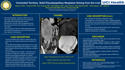
Introduction: Solid pseudopapillary neoplasms (SPN) tend to be rare, large, solid and cystic tumors of the pancreas that predominantly affect younger women. Although rare, common sites of metastases are the liver and local invasion to the duodenum, portal vein, and spleen. Here, we report a case of an SPN originating from the liver instead of the pancreas, which has not previously been reported in the literature.
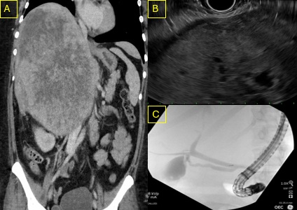
Case Description/Methods: This is an 18-year-old female with a history of discoid lupus who initially presented to an outside hospital with acute abdominal pain. CT and MRI demonstrated a heterogenous 25 cm mass arising from the right hepatic lobe (Image A). CT guided biopsy showed central necrosis and histology consistent with a SPN. She was then admitted to our hospital 1 month later due to symptoms of weight loss, vomiting, and obstructive jaundice with elevated total bilirubin (5.0 mg/dL). Subsequent EUS/ERCP revealed a massive liver tumor (Image B), a long left intrahepatic stricture and a non-dilated common bile duct (Image C). A 10 Fr x 18 cm biliary stent was placed for biliary decompression. Despite improved bilirubin level after stenting, the patient continued to have intermittent abdominal pain. Thus, she underwent an exploratory laparotomy, open cholecystectomy, and intraoperative liver biopsy. Whipple procedure was aborted due to intraoperative findings which clearly identified that the tumor was confined to the liver and did not originate from the pancreas. Liver biopsies showed evidence of stage 2/4 periportal fibrosis with moderate macrovesicular steatosis. Biopsies of the liver mass showed preliminary results of neoplastic tissue, but final tumor classification is still pending.
Discussion: In our review of the literature, SPNs are most associated with and defined as being of pancreatic origin. To our knowledge, this is the first case of a SPN of liver origin. Despite multiple imaging modalities including CT, MRI, and EUS as well as open exploratory surgery, a lesion was not identified within the pancreas. Although SPNs occur most often in younger women, this patient’s age at presentation of 18 was significantly younger than the mean age of 28.5. Due to the favorable prognosis, surgical resection remains the mainstay of treatment. This case draws attention to the enigmatic nature of SPNs and warrants further investigation.

Disclosures:
Bryant Le, MD1, Yousif Arif, BS2, David L. Cheung, MD3, Nabil El Hage Chehade, MD4, Brian Mendoza, MD3, Joshua Kwon, MD1, Peter H. Nguyen, MD3, Jason Samarasena, MD3, Frances Dang, MD3. P4702 - Uncharted Territory: Solid Pseudopapillary Neoplasm Arising from the Liver, ACG 2024 Annual Scientific Meeting Abstracts. Philadelphia, PA: American College of Gastroenterology.
1University of California Irvine, Orange, CA; 2University of California Irvine School of Medicine, Orange, CA; 3University of California, Irvine, Orange, CA; 4Scripps Clinic, San Diego, CA
Introduction: Solid pseudopapillary neoplasms (SPN) tend to be rare, large, solid and cystic tumors of the pancreas that predominantly affect younger women. Although rare, common sites of metastases are the liver and local invasion to the duodenum, portal vein, and spleen. Here, we report a case of an SPN originating from the liver instead of the pancreas, which has not previously been reported in the literature.
Case Description/Methods: This is an 18-year-old female with a history of discoid lupus who initially presented to an outside hospital with acute abdominal pain. CT and MRI demonstrated a heterogenous 25 cm mass arising from the right hepatic lobe (Image A). CT guided biopsy showed central necrosis and histology consistent with a SPN. She was then admitted to our hospital 1 month later due to symptoms of weight loss, vomiting, and obstructive jaundice with elevated total bilirubin (5.0 mg/dL). Subsequent EUS/ERCP revealed a massive liver tumor (Image B), a long left intrahepatic stricture and a non-dilated common bile duct (Image C). A 10 Fr x 18 cm biliary stent was placed for biliary decompression. Despite improved bilirubin level after stenting, the patient continued to have intermittent abdominal pain. Thus, she underwent an exploratory laparotomy, open cholecystectomy, and intraoperative liver biopsy. Whipple procedure was aborted due to intraoperative findings which clearly identified that the tumor was confined to the liver and did not originate from the pancreas. Liver biopsies showed evidence of stage 2/4 periportal fibrosis with moderate macrovesicular steatosis. Biopsies of the liver mass showed preliminary results of neoplastic tissue, but final tumor classification is still pending.
Discussion: In our review of the literature, SPNs are most associated with and defined as being of pancreatic origin. To our knowledge, this is the first case of a SPN of liver origin. Despite multiple imaging modalities including CT, MRI, and EUS as well as open exploratory surgery, a lesion was not identified within the pancreas. Although SPNs occur most often in younger women, this patient’s age at presentation of 18 was significantly younger than the mean age of 28.5. Due to the favorable prognosis, surgical resection remains the mainstay of treatment. This case draws attention to the enigmatic nature of SPNs and warrants further investigation.

Figure: Image A: Heterogenous liver mass on initial CT A/P
Image B: Large liver tumor noted on EUS
Image C: ERCP fluoroscopy showing intrahepatic stricture
Image B: Large liver tumor noted on EUS
Image C: ERCP fluoroscopy showing intrahepatic stricture
Disclosures:
Bryant Le indicated no relevant financial relationships.
Yousif Arif indicated no relevant financial relationships.
David Cheung indicated no relevant financial relationships.
Nabil El Hage Chehade indicated no relevant financial relationships.
Brian Mendoza indicated no relevant financial relationships.
Joshua Kwon indicated no relevant financial relationships.
Peter Nguyen indicated no relevant financial relationships.
Jason Samarasena: Cook Medical – Consultant. Medtronic – Advisory Committee/Board Member, Consultant. Neptune Medical – Advisory Committee/Board Member, Consultant. Olympus – Advisory Committee/Board Member, Consultant. Ovesco – Advisory Committee/Board Member, Consultant, Speakers Bureau. SatisfAI – Stock-privately held company. Steris – Advisory Committee/Board Member.
Frances Dang indicated no relevant financial relationships.
Bryant Le, MD1, Yousif Arif, BS2, David L. Cheung, MD3, Nabil El Hage Chehade, MD4, Brian Mendoza, MD3, Joshua Kwon, MD1, Peter H. Nguyen, MD3, Jason Samarasena, MD3, Frances Dang, MD3. P4702 - Uncharted Territory: Solid Pseudopapillary Neoplasm Arising from the Liver, ACG 2024 Annual Scientific Meeting Abstracts. Philadelphia, PA: American College of Gastroenterology.
