Tuesday Poster Session
Category: Liver
P4717 - Cardiac Clot or Cancer? A Presentation of Metastatic HCC
Tuesday, October 29, 2024
10:30 AM - 4:00 PM ET
Location: Exhibit Hall E

Has Audio

Kyle Hamann, DO
Riverside Medical Center
Kankakee, IL
Presenting Author(s)
Award: Presidential Poster Award
Kyle Hamann, DO1, Vijay Kata, MD2, Dhruv Joshi, DO1
1Riverside Medical Center, Kankakee, IL; 2Riverside Medical Group, Bourbonnais, IL
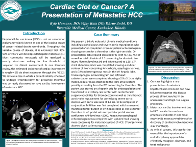
Introduction: Hepatocellular carcinoma (HCC) is not an uncommon malignancy widely known as one of the leading causes of cancer related deaths worldwide. Throughout the variable course of disease, it is estimated that 30%-50% of HCC’s will develop extrahepatic metastasis. Most commonly, metastasis will be restricted to nearby structures lowering the threshold of suspicion for distant involvement. The estimated incidence of cardiac involvement is roughly 6% via direct extension through the IVC. We review a case in which a patient initially scheduled to undergo thrombectomy for presumed thrombus was correctly discovered to have cardiac involvement of metastatic HCC.
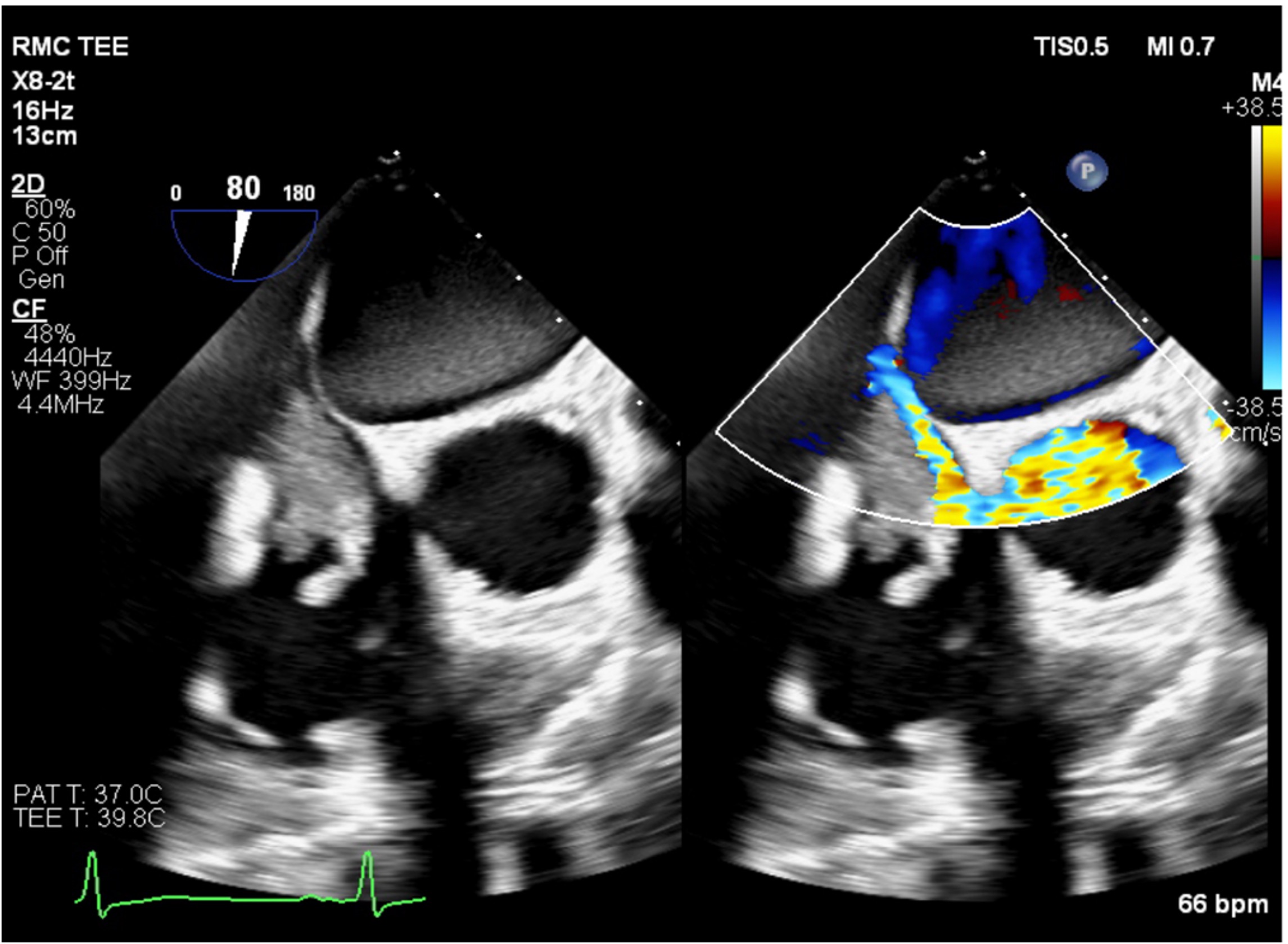
Case Description/Methods: We present a 66 y/o male with chronic alcoholism and severe aortic regurgitation who presented after completion of an outpatient echocardiogram showing concern for right ventricular thrombus. Initial labs showed transaminitis with ALT 30, AST 69 and Total bilirubin 1.6 consistent with alcoholic liver injury. Platelet level was 96 and INR elevated to 1.25. CTA chest and abdomen was completed showing a nodular liver concerning for cirrhosis, esophageal varices, and a 3.9 cm heterogenous mass in the left hepatic lobe. Transesophageal echocardiogram was completed showing a 2.9 x 5.1 cm highly mobile mass attached to the intra-atrial septum extending from the IVC concerning for thrombus. The patient was started on a heparin drip for anticoagulation and transferred to a tertiary care center for thrombectomy as well as transthoracic aortic valve replacement due to severe aortic stenosis with aortic valve area of 1.1 cm. MRI liver completed which uncovered multifocal tumor burden in left hepatic lobe and tumor thrombus in left portal vein and perihilar portal venous confluence. AFP level was >2000. Repeat transesophageal echocardiogram was completed with updated read showing mass concerning for metastatic spread of HCC. CT surgery was aborted given prognosis.
Discussion: Our case highlights a rare presentation of metastatic carcinoma and how failure to recognize this disease process almost resulted in an unnecessary and high-risk surgical procedure. It is important to understand cardiac involvement of HCC can be used as a prognostic indicator. In one small study(n=8), mean survival time after discovery of cardiac involvement was 3.67 months. As with all cancers, this case further exemplifies the importance of a multidisciplinary approach to effectively recognize, diagnose, and treat malignancy.

Disclosures:
Kyle Hamann, DO1, Vijay Kata, MD2, Dhruv Joshi, DO1. P4717 - Cardiac Clot or Cancer? A Presentation of Metastatic HCC, ACG 2024 Annual Scientific Meeting Abstracts. Philadelphia, PA: American College of Gastroenterology.
Kyle Hamann, DO1, Vijay Kata, MD2, Dhruv Joshi, DO1
1Riverside Medical Center, Kankakee, IL; 2Riverside Medical Group, Bourbonnais, IL
Introduction: Hepatocellular carcinoma (HCC) is not an uncommon malignancy widely known as one of the leading causes of cancer related deaths worldwide. Throughout the variable course of disease, it is estimated that 30%-50% of HCC’s will develop extrahepatic metastasis. Most commonly, metastasis will be restricted to nearby structures lowering the threshold of suspicion for distant involvement. The estimated incidence of cardiac involvement is roughly 6% via direct extension through the IVC. We review a case in which a patient initially scheduled to undergo thrombectomy for presumed thrombus was correctly discovered to have cardiac involvement of metastatic HCC.
Case Description/Methods: We present a 66 y/o male with chronic alcoholism and severe aortic regurgitation who presented after completion of an outpatient echocardiogram showing concern for right ventricular thrombus. Initial labs showed transaminitis with ALT 30, AST 69 and Total bilirubin 1.6 consistent with alcoholic liver injury. Platelet level was 96 and INR elevated to 1.25. CTA chest and abdomen was completed showing a nodular liver concerning for cirrhosis, esophageal varices, and a 3.9 cm heterogenous mass in the left hepatic lobe. Transesophageal echocardiogram was completed showing a 2.9 x 5.1 cm highly mobile mass attached to the intra-atrial septum extending from the IVC concerning for thrombus. The patient was started on a heparin drip for anticoagulation and transferred to a tertiary care center for thrombectomy as well as transthoracic aortic valve replacement due to severe aortic stenosis with aortic valve area of 1.1 cm. MRI liver completed which uncovered multifocal tumor burden in left hepatic lobe and tumor thrombus in left portal vein and perihilar portal venous confluence. AFP level was >2000. Repeat transesophageal echocardiogram was completed with updated read showing mass concerning for metastatic spread of HCC. CT surgery was aborted given prognosis.
Discussion: Our case highlights a rare presentation of metastatic carcinoma and how failure to recognize this disease process almost resulted in an unnecessary and high-risk surgical procedure. It is important to understand cardiac involvement of HCC can be used as a prognostic indicator. In one small study(n=8), mean survival time after discovery of cardiac involvement was 3.67 months. As with all cancers, this case further exemplifies the importance of a multidisciplinary approach to effectively recognize, diagnose, and treat malignancy.

Figure: Figure 1: A large multi-lobular and highly mobile thrombus or mass (measured 2.5cm x 5.0 cm) possible extended from the IVC and somewhat partially attached to the interatrial septum
Disclosures:
Kyle Hamann indicated no relevant financial relationships.
Vijay Kata indicated no relevant financial relationships.
Dhruv Joshi indicated no relevant financial relationships.
Kyle Hamann, DO1, Vijay Kata, MD2, Dhruv Joshi, DO1. P4717 - Cardiac Clot or Cancer? A Presentation of Metastatic HCC, ACG 2024 Annual Scientific Meeting Abstracts. Philadelphia, PA: American College of Gastroenterology.

