Tuesday Poster Session
Category: Liver
P4768 - ESBL E. coli Growth in a Multiloculated Liver Collection in an Elderly Patient With Prior Echinococcus and Ameba Infections - Treatment Dilemma
Tuesday, October 29, 2024
10:30 AM - 4:00 PM ET
Location: Exhibit Hall E

Has Audio
- IJ
Ihab Jameel, MD
NYC Health + Hospitals/Harlem
New York, NY
Presenting Author(s)
Ihab Jameel, MD, Mohamed K.. Osman, MD, MPH, Fnu Allahyar, MD, Joan Culpepper-Morgan, MD, FACG
NYC Health + Hospitals/Harlem, New York, NY
Introduction: A liver abscess is a pus-filled mass in the liver that can develop from injury to the liver or from an intra-abdominal infection. The majority of abscesses are either pyogenic or amebic. Pyogenic liver abscesses (PLA) are usually polymicrobial and present with life-threatening signs of sepsis. Here, we describe an atypical presentation of a pyogenic liver abscess in an elderly female with positive Echinococcus and E. histolytica serology.
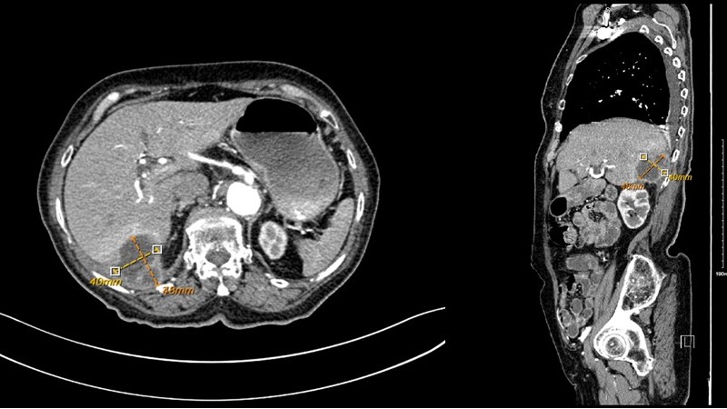
Case Description/Methods: A 96-year-old female with a history of hypertension, diverticulosis, laparoscopic cholecystectomy, and ERCP 3 years ago for choledocholithiasis, presented with chest pain, epigastric pain and shortness of breath. She had an associated episode of non-bloody vomiting and two episodes of diarrhea. The patient frequently traveled between the Dominican Republic and the USA. Initial examination revealed stable vital signs, clear chest, normal heart sounds, soft non-tender abdomen, and present bowel sounds. The neurological exam was non-focal. Laboratory investigations showed normal leukocyte count, kidney function, liver function, and electrolytes. Troponins and EKG were unremarkable. CT Chest/abdomen, to rule out aortic dissection, revealed a rim-enhancing, multiloculated cystic mass arising from the sub-diaphragmatic posterior right hepatic lobe, along with a small reactive right-sided pleural effusion. MRI confirmed it to be a multilocular abscess. Serology positive for E. histolytica and Echinococcus prompted initial therapy with metronidazole and albendazole. Diagnostic, ultrasound-guided cyst aspiration and culture revealed few WBCs and few ESBL E. Coli, for which the patient was started on IV meropenem. A week later, CT-guided drainage of the hepatic collection via transhepatic route, was performed with placement of a pigtail catheter. This was followed by a spike in WBC from 8.12 x10³/uL to 35.4 x10³/uL; an absolute eosinophil spike from 0.43 to 4.05 K/uL; and a fever spike that improved with antibiotic therapy. The patient was discharged with long-term IV antibiotics.
Discussion: Both echinococcal and amebic infections of the liver can be quiescent for years. We suspect that our patient had a latent echinococcal cyst that became secondarily infected with ESBL E.Coli. However, our patient was notably afebrile with a normal white count until the drain was placed. Then she had a fever, leukocytosis, and eosinophilia suggesting systemic release of parasitic material. Blood cultures were negative. Pain was her main symptom.

Disclosures:
Ihab Jameel, MD, Mohamed K.. Osman, MD, MPH, Fnu Allahyar, MD, Joan Culpepper-Morgan, MD, FACG. P4768 - ESBL <i>E. coli</i> Growth in a Multiloculated Liver Collection in an Elderly Patient With Prior Echinococcus and Ameba Infections - Treatment Dilemma, ACG 2024 Annual Scientific Meeting Abstracts. Philadelphia, PA: American College of Gastroenterology.
NYC Health + Hospitals/Harlem, New York, NY
Introduction: A liver abscess is a pus-filled mass in the liver that can develop from injury to the liver or from an intra-abdominal infection. The majority of abscesses are either pyogenic or amebic. Pyogenic liver abscesses (PLA) are usually polymicrobial and present with life-threatening signs of sepsis. Here, we describe an atypical presentation of a pyogenic liver abscess in an elderly female with positive Echinococcus and E. histolytica serology.
Case Description/Methods: A 96-year-old female with a history of hypertension, diverticulosis, laparoscopic cholecystectomy, and ERCP 3 years ago for choledocholithiasis, presented with chest pain, epigastric pain and shortness of breath. She had an associated episode of non-bloody vomiting and two episodes of diarrhea. The patient frequently traveled between the Dominican Republic and the USA. Initial examination revealed stable vital signs, clear chest, normal heart sounds, soft non-tender abdomen, and present bowel sounds. The neurological exam was non-focal. Laboratory investigations showed normal leukocyte count, kidney function, liver function, and electrolytes. Troponins and EKG were unremarkable. CT Chest/abdomen, to rule out aortic dissection, revealed a rim-enhancing, multiloculated cystic mass arising from the sub-diaphragmatic posterior right hepatic lobe, along with a small reactive right-sided pleural effusion. MRI confirmed it to be a multilocular abscess. Serology positive for E. histolytica and Echinococcus prompted initial therapy with metronidazole and albendazole. Diagnostic, ultrasound-guided cyst aspiration and culture revealed few WBCs and few ESBL E. Coli, for which the patient was started on IV meropenem. A week later, CT-guided drainage of the hepatic collection via transhepatic route, was performed with placement of a pigtail catheter. This was followed by a spike in WBC from 8.12 x10³/uL to 35.4 x10³/uL; an absolute eosinophil spike from 0.43 to 4.05 K/uL; and a fever spike that improved with antibiotic therapy. The patient was discharged with long-term IV antibiotics.
Discussion: Both echinococcal and amebic infections of the liver can be quiescent for years. We suspect that our patient had a latent echinococcal cyst that became secondarily infected with ESBL E.Coli. However, our patient was notably afebrile with a normal white count until the drain was placed. Then she had a fever, leukocytosis, and eosinophilia suggesting systemic release of parasitic material. Blood cultures were negative. Pain was her main symptom.

Figure: Axial and sagittal view of CT Abdomen and Pelvis scan with IV contrast showing a 4.8 x 4.0 cm rim enhancing, multiloculated, cystic mass arising from the subdiaphragmatic posterior right hepatic lobe.
Disclosures:
Ihab Jameel indicated no relevant financial relationships.
Mohamed Osman indicated no relevant financial relationships.
Fnu Allahyar indicated no relevant financial relationships.
Joan Culpepper-Morgan indicated no relevant financial relationships.
Ihab Jameel, MD, Mohamed K.. Osman, MD, MPH, Fnu Allahyar, MD, Joan Culpepper-Morgan, MD, FACG. P4768 - ESBL <i>E. coli</i> Growth in a Multiloculated Liver Collection in an Elderly Patient With Prior Echinococcus and Ameba Infections - Treatment Dilemma, ACG 2024 Annual Scientific Meeting Abstracts. Philadelphia, PA: American College of Gastroenterology.
