Tuesday Poster Session
Category: Liver
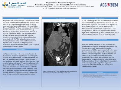
P4779 - Polycystic Liver Disease's Silent Squeeze: Unmasking Tachycardia - A Case Report and Review of the Literature
Tuesday, October 29, 2024
10:30 AM - 4:00 PM ET
Location: Exhibit Hall E

Has Audio

Nida Ansari, DO
St. Joseph's University Medical Center
Flanders, NJ
Presenting Author(s)
Nida Ansari, DO1, Sacide S. Ozgur, MD2, Gabriel Melki, MD2, Alisa Farokhian, MD2, Walid Baddoura, MD2
1St. Joseph's University Medical Center, Flanders, NJ; 2St. Joseph's University Medical Center, Paterson, NJ
Introduction: Polycystic Liver Disease (PLD) is a rare inherited disease due to the mutation of liver epithelial cells, allowing liver tissue to be replaced with fluid-filled cysts. Often associated with Autosomal Dominant Polycystic Kidney Disease (ADPKD), PLD can exist independently, likely a result of embryonic ductal plate malformation. Patients are asymptomatic, with incidental detection on CT scan. Patients can present with symptoms of mass effect on adjacent structures, especially vascular structures, causing portal hypertension and associated complications. Although cardiac compression resulting from mass effect is rare, with fewer than ten reported cases, we present an intriguing case of tachycardia in a patient with severe PLD, likely caused by compression of the right atrium.
Case Description/Methods: An 82 YO woman with a medical history of hypertension, pulmonary hypertension, cardiomyopathy, diastolic HF, hypothyroidism, and PLD presented to the ED with worsening bilateral lower extremity edema for two weeks and right upper quadrant abdominal pain. Labs revealed leukocytosis and a CT scan showed a large right pleural effusion for which she had a thoracentesis with chest tube placement. Due to hemodynamic instability and Enterobacter bacteremia, she was transferred to the ICU. Her course was complicated with tachycardia and multiple episodes of melena, resulting in a hemoglobin drop. GI was consulted for endoscopic intervention. A non-bleeding gastric and duodenal ulcer was found and treated with clips. Her melena resolved and her hemoglobin improved. She continued to experience tachycardia despite non-cardiac interventions. Multiple EKGs revealed sinus tachycardia. A TEE was performed, which was significant for external right atrial compression by left-sided liver cysts, and it was concluded to be the cause of her tachycardia.
Discussion: I
While it is acknowledged that PLD is often associated with external compression of surrounding structures, the literature at present is scant concerning cardiac compression. A PubMed search yielded only a few cases. Our case is noteworthy because, after eliminating other potential causes of tachycardia, we discovered that the patient's right atrium was externally compressed. Although her compression did not cause obstructive shock, external compression has the potential for more fatal complications. We aim to highlight this association's rarity and emphasize the importance of prompt recognition and treatment of PLD in patients with compression symptoms.

Disclosures:
Nida Ansari, DO1, Sacide S. Ozgur, MD2, Gabriel Melki, MD2, Alisa Farokhian, MD2, Walid Baddoura, MD2. P4779 - Polycystic Liver Disease's Silent Squeeze: Unmasking Tachycardia - A Case Report and Review of the Literature, ACG 2024 Annual Scientific Meeting Abstracts. Philadelphia, PA: American College of Gastroenterology.
1St. Joseph's University Medical Center, Flanders, NJ; 2St. Joseph's University Medical Center, Paterson, NJ
Introduction: Polycystic Liver Disease (PLD) is a rare inherited disease due to the mutation of liver epithelial cells, allowing liver tissue to be replaced with fluid-filled cysts. Often associated with Autosomal Dominant Polycystic Kidney Disease (ADPKD), PLD can exist independently, likely a result of embryonic ductal plate malformation. Patients are asymptomatic, with incidental detection on CT scan. Patients can present with symptoms of mass effect on adjacent structures, especially vascular structures, causing portal hypertension and associated complications. Although cardiac compression resulting from mass effect is rare, with fewer than ten reported cases, we present an intriguing case of tachycardia in a patient with severe PLD, likely caused by compression of the right atrium.
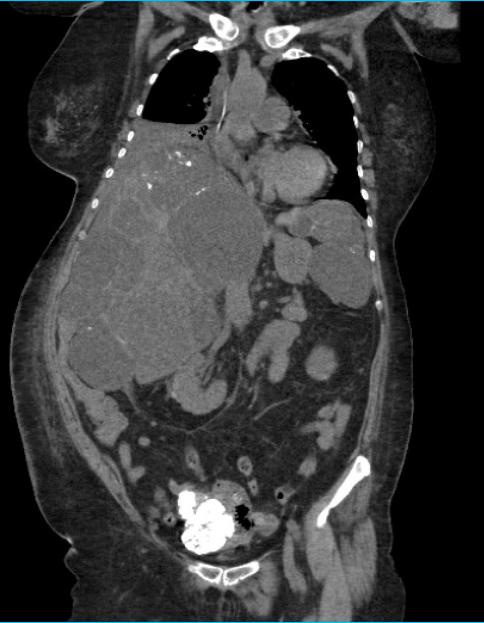
Case Description/Methods: An 82 YO woman with a medical history of hypertension, pulmonary hypertension, cardiomyopathy, diastolic HF, hypothyroidism, and PLD presented to the ED with worsening bilateral lower extremity edema for two weeks and right upper quadrant abdominal pain. Labs revealed leukocytosis and a CT scan showed a large right pleural effusion for which she had a thoracentesis with chest tube placement. Due to hemodynamic instability and Enterobacter bacteremia, she was transferred to the ICU. Her course was complicated with tachycardia and multiple episodes of melena, resulting in a hemoglobin drop. GI was consulted for endoscopic intervention. A non-bleeding gastric and duodenal ulcer was found and treated with clips. Her melena resolved and her hemoglobin improved. She continued to experience tachycardia despite non-cardiac interventions. Multiple EKGs revealed sinus tachycardia. A TEE was performed, which was significant for external right atrial compression by left-sided liver cysts, and it was concluded to be the cause of her tachycardia.
Discussion: I
While it is acknowledged that PLD is often associated with external compression of surrounding structures, the literature at present is scant concerning cardiac compression. A PubMed search yielded only a few cases. Our case is noteworthy because, after eliminating other potential causes of tachycardia, we discovered that the patient's right atrium was externally compressed. Although her compression did not cause obstructive shock, external compression has the potential for more fatal complications. We aim to highlight this association's rarity and emphasize the importance of prompt recognition and treatment of PLD in patients with compression symptoms.

Figure: Computer Tomography Abdomen and Pelvis, coronal view demonstrating extensive hepatic cysts that is externally compressing the right atrium.
Disclosures:
Nida Ansari indicated no relevant financial relationships.
Sacide Ozgur indicated no relevant financial relationships.
Gabriel Melki indicated no relevant financial relationships.
Alisa Farokhian indicated no relevant financial relationships.
Walid Baddoura indicated no relevant financial relationships.
Nida Ansari, DO1, Sacide S. Ozgur, MD2, Gabriel Melki, MD2, Alisa Farokhian, MD2, Walid Baddoura, MD2. P4779 - Polycystic Liver Disease's Silent Squeeze: Unmasking Tachycardia - A Case Report and Review of the Literature, ACG 2024 Annual Scientific Meeting Abstracts. Philadelphia, PA: American College of Gastroenterology.
