Tuesday Poster Session
Category: Liver
P4846 - Non-Thrombotic Budd-Chiari Syndrome Secondary to Significant Hepatomegaly
Tuesday, October 29, 2024
10:30 AM - 4:00 PM ET
Location: Exhibit Hall E

Has Audio

Hannah Murawsky, BS
Kirk Kerkorian School of Medicine at the University of Nevada
Las Vegas, NV
Presenting Author(s)
Hannah Murawsky, BS1, Amrit Narwan, MD1, Taha Shaikh, DO1, Rajan Amin, MD1, Vignan Manne, MD2
1Kirk Kerkorian School of Medicine at the University of Nevada, Las Vegas, NV; 2Kirk Kirkorian School of Medicine, Las Vegas, NV
Introduction: Budd Chiari Syndrome (BCS) is a rare congestive hepatopathy, most commonly caused by venous outflow obstruction secondary to thrombosis. Patients often present with abrupt onset of ascites and abdominal pain, and left untreated, it can progress to fulminant liver failure. Here, we report a case of BCS characterized by hepatic vein occlusion likely secondary to significant hepatomegaly with no evidence of thrombus.
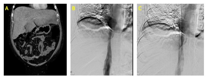
Case Description/Methods: A 28-year-old male with a history of alcohol use disorder presented with 2 weeks of upper abdominal pain and jaundice. Physical examination on admission was notable for scleral icterus, abdominal distention, and tenderness to palpation in the right upper quadrant. Laboratory values were notable for alkaline phosphatase 464, AST 179, ALT 50, total bilirubin 9.5, direct bilirubin 6.9, and GGT 942. Further studies including HIV, Hepatitis A, B, and C serologies, AFP, AMA, CMV, Antithrombin III, and Jak 2 mutation were negative or within normal limits. CT abdomen revealed mass effect causing diameter reduction of the inferior vena cava (IVC) by 50%. MRI abdomen revealed hepatomegaly measuring 20cm, enlarged caudate lobe, narrow caliber of the IVC and hepatic veins consistent with BCS; of note, no thrombus was seen on imaging. He then underwent venography of the IVC, right and middle hepatic veins which revealed significant stenosis, status post balloon angioplasty with near resolution of the stenosis.
Discussion: The vast majority of BCS presents as hepatic vein obstruction due to thrombus, often in the setting of hypercoagulability. Rarely, adjacent masses, cysts, or abscesses can cause narrowing of the hepatic veins by mass effect. Here we present a case of hepatic venous outflow obstruction likely secondary to extrinsic compression in the setting of hepatomegaly. This presentation suggests that BCS should still be considered when there is concern for hepatic outflow obstruction, even without evidence of thrombus or mass.

Disclosures:
Hannah Murawsky, BS1, Amrit Narwan, MD1, Taha Shaikh, DO1, Rajan Amin, MD1, Vignan Manne, MD2. P4846 - Non-Thrombotic Budd-Chiari Syndrome Secondary to Significant Hepatomegaly, ACG 2024 Annual Scientific Meeting Abstracts. Philadelphia, PA: American College of Gastroenterology.
1Kirk Kerkorian School of Medicine at the University of Nevada, Las Vegas, NV; 2Kirk Kirkorian School of Medicine, Las Vegas, NV
Introduction: Budd Chiari Syndrome (BCS) is a rare congestive hepatopathy, most commonly caused by venous outflow obstruction secondary to thrombosis. Patients often present with abrupt onset of ascites and abdominal pain, and left untreated, it can progress to fulminant liver failure. Here, we report a case of BCS characterized by hepatic vein occlusion likely secondary to significant hepatomegaly with no evidence of thrombus.
Case Description/Methods: A 28-year-old male with a history of alcohol use disorder presented with 2 weeks of upper abdominal pain and jaundice. Physical examination on admission was notable for scleral icterus, abdominal distention, and tenderness to palpation in the right upper quadrant. Laboratory values were notable for alkaline phosphatase 464, AST 179, ALT 50, total bilirubin 9.5, direct bilirubin 6.9, and GGT 942. Further studies including HIV, Hepatitis A, B, and C serologies, AFP, AMA, CMV, Antithrombin III, and Jak 2 mutation were negative or within normal limits. CT abdomen revealed mass effect causing diameter reduction of the inferior vena cava (IVC) by 50%. MRI abdomen revealed hepatomegaly measuring 20cm, enlarged caudate lobe, narrow caliber of the IVC and hepatic veins consistent with BCS; of note, no thrombus was seen on imaging. He then underwent venography of the IVC, right and middle hepatic veins which revealed significant stenosis, status post balloon angioplasty with near resolution of the stenosis.
Discussion: The vast majority of BCS presents as hepatic vein obstruction due to thrombus, often in the setting of hypercoagulability. Rarely, adjacent masses, cysts, or abscesses can cause narrowing of the hepatic veins by mass effect. Here we present a case of hepatic venous outflow obstruction likely secondary to extrinsic compression in the setting of hepatomegaly. This presentation suggests that BCS should still be considered when there is concern for hepatic outflow obstruction, even without evidence of thrombus or mass.

Figure: Figure 1: CT Abdomen Pelvis and IR Venography. Figure 1A: CTAP with contrast demonstrating significant hepatomegaly. Figure 1B: IR venography demonstrating significant stenosis of the IVC. Figure 1C: IR venography showing IVC post-dilation.
Disclosures:
Hannah Murawsky indicated no relevant financial relationships.
Amrit Narwan indicated no relevant financial relationships.
Taha Shaikh indicated no relevant financial relationships.
Rajan Amin indicated no relevant financial relationships.
Vignan Manne indicated no relevant financial relationships.
Hannah Murawsky, BS1, Amrit Narwan, MD1, Taha Shaikh, DO1, Rajan Amin, MD1, Vignan Manne, MD2. P4846 - Non-Thrombotic Budd-Chiari Syndrome Secondary to Significant Hepatomegaly, ACG 2024 Annual Scientific Meeting Abstracts. Philadelphia, PA: American College of Gastroenterology.
