Tuesday Poster Session
Category: Liver
P4865 - Sarcomatoid HCC: The Freak of TACE
Tuesday, October 29, 2024
10:30 AM - 4:00 PM ET
Location: Exhibit Hall E

Has Audio

Oana Vester, MD
The Ohio State University Wexner Medical Center
Columbus, OH
Presenting Author(s)
Oana Vester, MD, Chathur Acharya, MBBS, Vivek Mendiratta, MD
The Ohio State University Wexner Medical Center, Columbus, OH
Introduction: Hepatocellular carcinoma (HCC) is the 6th most common cause of death in the United States. Among subtypes of HCC, sarcomatoid HCC is one of the most rare and aggressive subtypes. Here we present a rare case of sarcomatoid HCC diagnosed on liver explanation after transarterial chemoembolization (TACE) and its chronology of progression after liver transplant.
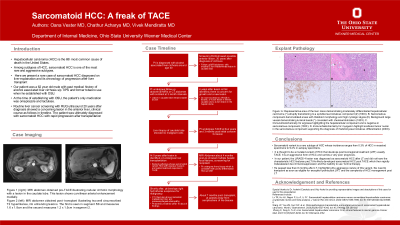
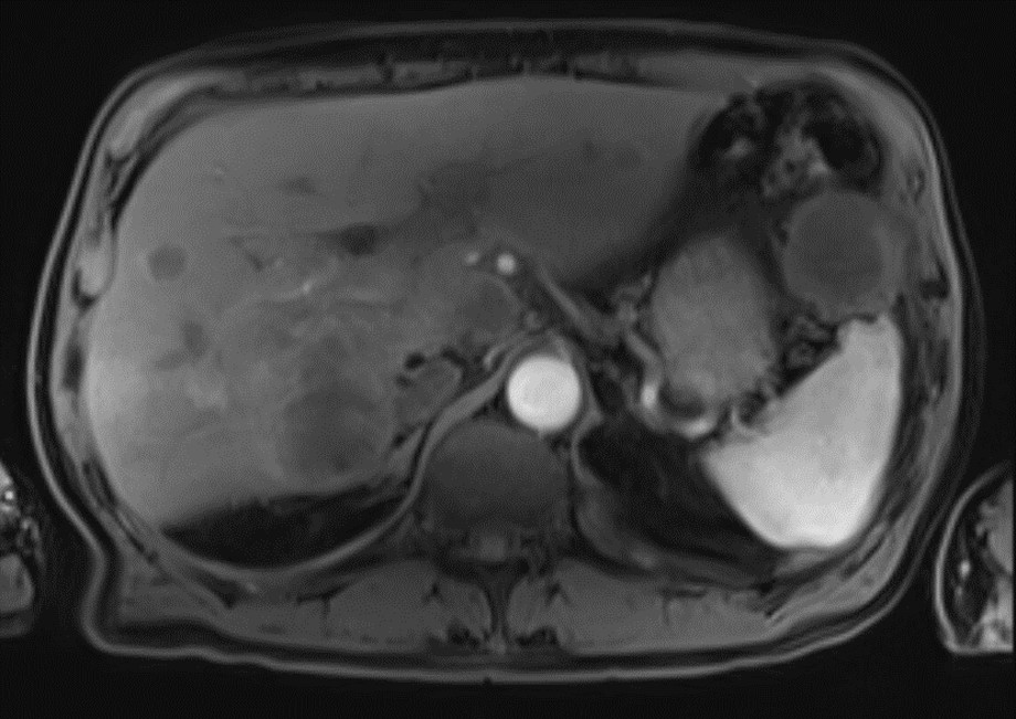
Case Description/Methods: Our patient was a 62 year old male with alcohol associated cirrhosis. Routine liver cancer screening with RUQ ultrasound 20 years after diagnosis showed a concerning lesion in the anterior liver. Follow up MRI showed an exophytic 3cm lesion located in the inferior aspect of the caudate lobe without features concerning for HCC. Two years later, triple phase CT scan abdomen showed a 4.3cm LIRADS 4 lesion in the caudate and a new concerning 2.3cm LIRADS 4 central hepatic dome lesion. Liver biopsy of the caudate lobe mass via EUS showed no malignant cells. Interval MRI 3 months later showed a mild increase in size of the caudate lobe lesion to 4.6cm. After discussion in tumor board, the patient underwent TACE with liver transplantation (LT) 8 months after. Liver explant pathology showed HCC, sarcomatoid variant (with reduced Cam 5.2 expression and diffusely positive Desmin) with partial treatment response and vascular invasion. Surveillance imaging with MRI 3 months post-LT was concerning for multifocal neoplasm in the liver as shown in Figure 1. CT chest imaging showed right hydrothorax suspicious for malignant effusion, and metastatic mediastinal, hilar, and paraoesophageal adenopathy. The patient started Lenvatinib within 10 days of these findings. The patient passed away from complications of his disease 2 weeks later.
Discussion: Sarcomatoid variant is a rare subtype of HCC whose incidence ranges from 0.3% of HCC in resected specimens to 9.4% in autopsy specimens. It is thought to be a mutated variant of HCC that develops post locoregional treatment (LRT), usually TACE. It is an aggressive form of HCC and carries a very poor prognosis. In our patient, the LiRADS 4 lesion was diagnosed as sarcomatoid HCC after LT and did not have the characteristic HCC features preLT. He likely developed sarcomatoid HCC post-TACE which then rapidly metastasized due immunosuppression and the inability to use 1st line therapy. He passed less than 6 months after LT, highlighting the aggressive nature of this variant, the need to transplant as soon as eligible for exception points post-LRT, and the complexity of HCC management post LT.

Disclosures:
Oana Vester, MD, Chathur Acharya, MBBS, Vivek Mendiratta, MD. P4865 - Sarcomatoid HCC: The Freak of TACE, ACG 2024 Annual Scientific Meeting Abstracts. Philadelphia, PA: American College of Gastroenterology.
The Ohio State University Wexner Medical Center, Columbus, OH
Introduction: Hepatocellular carcinoma (HCC) is the 6th most common cause of death in the United States. Among subtypes of HCC, sarcomatoid HCC is one of the most rare and aggressive subtypes. Here we present a rare case of sarcomatoid HCC diagnosed on liver explanation after transarterial chemoembolization (TACE) and its chronology of progression after liver transplant.
Case Description/Methods: Our patient was a 62 year old male with alcohol associated cirrhosis. Routine liver cancer screening with RUQ ultrasound 20 years after diagnosis showed a concerning lesion in the anterior liver. Follow up MRI showed an exophytic 3cm lesion located in the inferior aspect of the caudate lobe without features concerning for HCC. Two years later, triple phase CT scan abdomen showed a 4.3cm LIRADS 4 lesion in the caudate and a new concerning 2.3cm LIRADS 4 central hepatic dome lesion. Liver biopsy of the caudate lobe mass via EUS showed no malignant cells. Interval MRI 3 months later showed a mild increase in size of the caudate lobe lesion to 4.6cm. After discussion in tumor board, the patient underwent TACE with liver transplantation (LT) 8 months after. Liver explant pathology showed HCC, sarcomatoid variant (with reduced Cam 5.2 expression and diffusely positive Desmin) with partial treatment response and vascular invasion. Surveillance imaging with MRI 3 months post-LT was concerning for multifocal neoplasm in the liver as shown in Figure 1. CT chest imaging showed right hydrothorax suspicious for malignant effusion, and metastatic mediastinal, hilar, and paraoesophageal adenopathy. The patient started Lenvatinib within 10 days of these findings. The patient passed away from complications of his disease 2 weeks later.
Discussion: Sarcomatoid variant is a rare subtype of HCC whose incidence ranges from 0.3% of HCC in resected specimens to 9.4% in autopsy specimens. It is thought to be a mutated variant of HCC that develops post locoregional treatment (LRT), usually TACE. It is an aggressive form of HCC and carries a very poor prognosis. In our patient, the LiRADS 4 lesion was diagnosed as sarcomatoid HCC after LT and did not have the characteristic HCC features preLT. He likely developed sarcomatoid HCC post-TACE which then rapidly metastasized due immunosuppression and the inability to use 1st line therapy. He passed less than 6 months after LT, highlighting the aggressive nature of this variant, the need to transplant as soon as eligible for exception points post-LRT, and the complexity of HCC management post LT.

Figure: Figure 1: Staging MRI abdomen illustrating two well circumscribed T2 hyperintense, rim-enhancing lesions. The first is seen in segment 5/8 and measures 1.6 x 1.8cm and the second measures 1.2 x 1.5cm in segment 2.
Disclosures:
Oana Vester indicated no relevant financial relationships.
Chathur Acharya indicated no relevant financial relationships.
Vivek Mendiratta indicated no relevant financial relationships.
Oana Vester, MD, Chathur Acharya, MBBS, Vivek Mendiratta, MD. P4865 - Sarcomatoid HCC: The Freak of TACE, ACG 2024 Annual Scientific Meeting Abstracts. Philadelphia, PA: American College of Gastroenterology.
