Tuesday Poster Session
Category: Small Intestine
P4954 - Peritoneal Calcifications: A Rare Cause of Small Bowel Obstruction
Tuesday, October 29, 2024
10:30 AM - 4:00 PM ET
Location: Exhibit Hall E

Has Audio
.jpg)
Brandon Truong, MD
Cedars-Sinai Medical Center
Los Angeles, CA
Presenting Author(s)
Brandon Truong, MD1, Harman K. Rahal, MD1, Lester Tsai, MD1, Bianca W. Chang, MD1, Jane Lim, MD1, Ali Rezaie, MD2, Amrit K.. Kamboj, MD1
1Cedars-Sinai Medical Center, Los Angeles, CA; 2Cedars-Sinai Medical Center, West Hollywood, CA
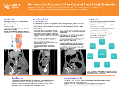
Introduction: Peritoneal calcifications are a rare finding seen on cross-sectional imaging and can occur secondary to various benign and neoplastic etiologies. Here, we highlight a patient case where peritoneal calcifications led to recurrent small bowel obstruction.
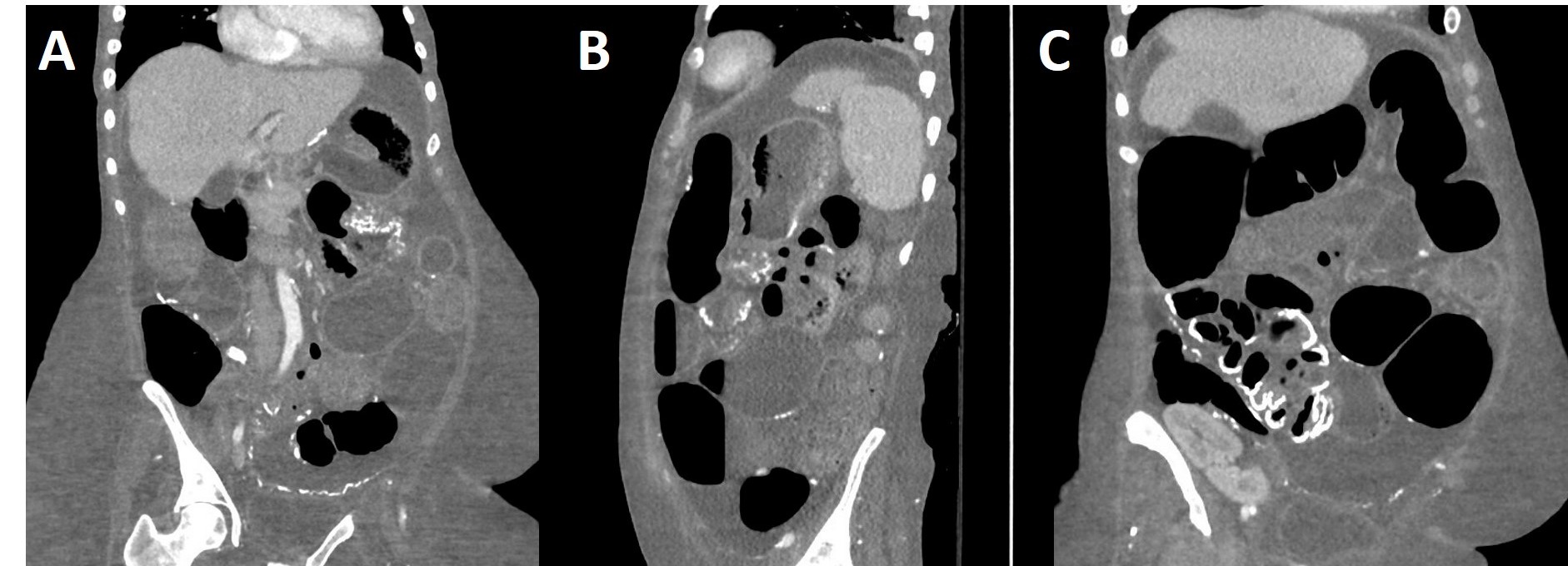
Case Description/Methods: A 59-year-old woman with a history of end-stage renal disease requiring peritoneal dialysis two years prior status-post kidney transplantation, renal cell carcinoma status-post right nephrectomy, and recurrent bacterial peritonitis presented with acute on chronic abdominal pain and inability to tolerate oral intake. Computed tomography (CT) abdomen revealed moderate ascites, extensive serosal calcifications with sclerosing peritonitis, and upstream dilatation of gastrointestinal tract consistent with partial small bowel obstruction (Figure). Paracentesis upon admission demonstrated spontaneous bacterial peritonitis (SBP) with white blood cell count of 6,954 with 87% neutrophils without evidence of malignancy. The patient was treated with broad spectrum antibiotics, underwent nasogastric tube decompression, and initiated on total parenteral nutrition. Unfortunately, the patient would struggle with prolonged symptomatic partial small bowel obstruction requiring continued nasogastric decompression. Patient was deemed to not be a candidate for surgical intervention due to the severity of calcifications and underlying comorbidities. The patient remained admitted for continued conservative management of obstruction, eventually improving enough to tolerate oral intake, and was subsequently discharged to a long-term acute care facility.
Discussion: Peritoneal calcifications can develop due to a breadth of etiologies including fat necrosis or infarcts calcifying over time, various infections ranging from parasites to tuberculosis, recurrent bacterial or chemical peritonitis, peritoneal dialysis, mineral imbalances like hypercalcemia, and intra-abdominal malignancies. Here, we highlight a patient case where peritoneal dialysis and recurrent bacterial peritonitis resulted in the development of extensive peritoneal calcifications. This case also highlights that peritoneal calcifications can rarely cause significant complications such as small bowel obstruction.

Disclosures:
Brandon Truong, MD1, Harman K. Rahal, MD1, Lester Tsai, MD1, Bianca W. Chang, MD1, Jane Lim, MD1, Ali Rezaie, MD2, Amrit K.. Kamboj, MD1. P4954 - Peritoneal Calcifications: A Rare Cause of Small Bowel Obstruction, ACG 2024 Annual Scientific Meeting Abstracts. Philadelphia, PA: American College of Gastroenterology.
1Cedars-Sinai Medical Center, Los Angeles, CA; 2Cedars-Sinai Medical Center, West Hollywood, CA
Introduction: Peritoneal calcifications are a rare finding seen on cross-sectional imaging and can occur secondary to various benign and neoplastic etiologies. Here, we highlight a patient case where peritoneal calcifications led to recurrent small bowel obstruction.
Case Description/Methods: A 59-year-old woman with a history of end-stage renal disease requiring peritoneal dialysis two years prior status-post kidney transplantation, renal cell carcinoma status-post right nephrectomy, and recurrent bacterial peritonitis presented with acute on chronic abdominal pain and inability to tolerate oral intake. Computed tomography (CT) abdomen revealed moderate ascites, extensive serosal calcifications with sclerosing peritonitis, and upstream dilatation of gastrointestinal tract consistent with partial small bowel obstruction (Figure). Paracentesis upon admission demonstrated spontaneous bacterial peritonitis (SBP) with white blood cell count of 6,954 with 87% neutrophils without evidence of malignancy. The patient was treated with broad spectrum antibiotics, underwent nasogastric tube decompression, and initiated on total parenteral nutrition. Unfortunately, the patient would struggle with prolonged symptomatic partial small bowel obstruction requiring continued nasogastric decompression. Patient was deemed to not be a candidate for surgical intervention due to the severity of calcifications and underlying comorbidities. The patient remained admitted for continued conservative management of obstruction, eventually improving enough to tolerate oral intake, and was subsequently discharged to a long-term acute care facility.
Discussion: Peritoneal calcifications can develop due to a breadth of etiologies including fat necrosis or infarcts calcifying over time, various infections ranging from parasites to tuberculosis, recurrent bacterial or chemical peritonitis, peritoneal dialysis, mineral imbalances like hypercalcemia, and intra-abdominal malignancies. Here, we highlight a patient case where peritoneal dialysis and recurrent bacterial peritonitis resulted in the development of extensive peritoneal calcifications. This case also highlights that peritoneal calcifications can rarely cause significant complications such as small bowel obstruction.

Figure: Figure: Computed tomography of the abdomen and pelvis (A) Coronal, (B) Sagittal, (C) Coronal, more posteriorly, showing small bowel distention with diffuse peritoneal calcifications.
Disclosures:
Brandon Truong indicated no relevant financial relationships.
Harman Rahal indicated no relevant financial relationships.
Lester Tsai indicated no relevant financial relationships.
Bianca Chang indicated no relevant financial relationships.
Jane Lim indicated no relevant financial relationships.
Ali Rezaie: Ardelyx – Consultant. Bausch Health – Consultant, Speakers Bureau. Gemelli Biotech – Stock-privately held company. GoodLFE – Stock-privately held company.
Amrit Kamboj: Castle Biosciences – Consultant.
Brandon Truong, MD1, Harman K. Rahal, MD1, Lester Tsai, MD1, Bianca W. Chang, MD1, Jane Lim, MD1, Ali Rezaie, MD2, Amrit K.. Kamboj, MD1. P4954 - Peritoneal Calcifications: A Rare Cause of Small Bowel Obstruction, ACG 2024 Annual Scientific Meeting Abstracts. Philadelphia, PA: American College of Gastroenterology.
