Tuesday Poster Session
Category: Small Intestine
P4958 - A Life-Threatening GI Bleed Secondary to Duodenal GIST Requiring Urgent Surgical Resection
Tuesday, October 29, 2024
10:30 AM - 4:00 PM ET
Location: Exhibit Hall E

Has Audio

Sanad Dawod, MD
Henry Ford Health
Detroit, MI
Presenting Author(s)
Sanad Dawod, MD1, Qais Dawod, MD2, Emily Smith, MD3, Carl V. Crawford, MD2, Rasa Zarnegar, MD2
1Henry Ford Health, Detroit, MI; 2New York-Presbyterian / Weill Cornell Medical Center, New York, NY; 3New York-Presbyterian/Weill Cornell, New York, NY
Introduction: Gastrointestinal stromal tumors (GISTs) are rare primary gastrointestinal neoplasms that can develop anywhere along the gastrointestinal (GI) tract. Presentation varies by location but can range from asymptomatic, to local obstructive symptoms and bleeding. They can form anywhere along the GI tract. Duodenal GIST is relatively rare when compared to other sites. We showcase a patient presenting with a life-threatening bleed secondary to duodenal GIST requiring surgical resection.
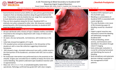
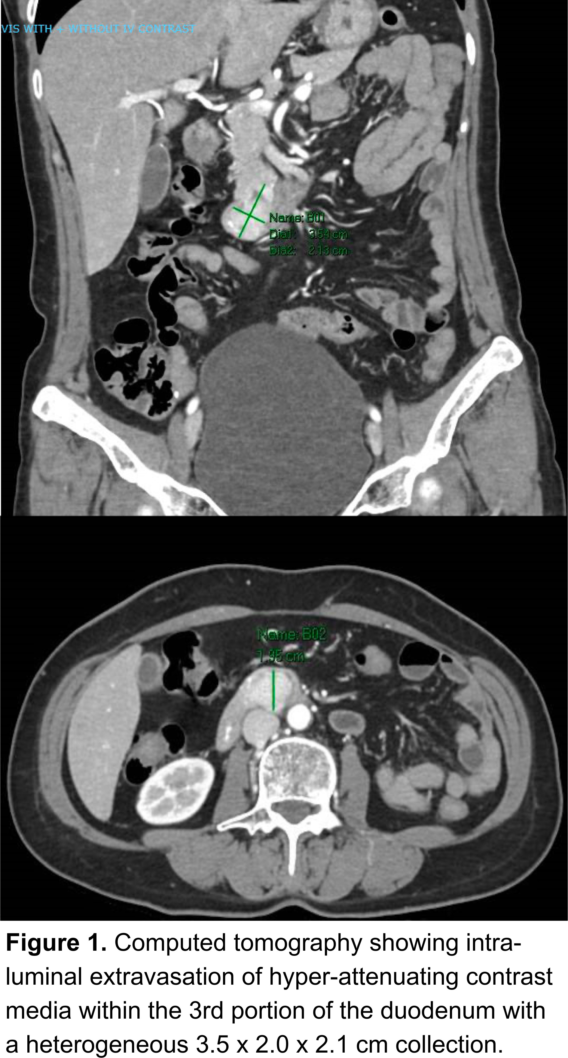
Case Description/Methods: A 64-year-old female patient with a past medical history of type 2 diabetes mellitus and gastro-esophageal reflux disease presented with three days of melenic stools, bilious emesis and left-sided abdominal pain. On exam, she was tachycardic and normotensive with tenderness to palpation in the left lower quadrant. Initial investigations revealed a hemoglobin 5.5 g/dl. She was resuscitated and a Computed Tomography Angiography (CTA) revealed active contrast intraluminal extravasation within the 3rd portion of duodenum with a mass-like collection of hyperattenuating material suggesting a intraluminal hematoma (Figure 1). The patient thereafter underwent an esophagoduodenoscopy (EGD) which showed a large, superficially ulcerated submucosal mass with a visible vessel in the third portion of the duodenum not actively bleeding. No intervention was performed due to close proximity to the ampulla. The endoscopic appearance was consistent with GIST. Surgery was consulted due to inability to biopsy the lesion and ensure adequate control of bleeding, and the patient underwent an open duodenal resection. The mass was measured at 3.9 x 2.1 x 1.7 cm. No lymphadenopathy was noted intra-operatively. Pathology confirmed a low-grade GIST with clean margins.
Discussion: Duodenal GISTs comprise up to 5% of all GIST occurrences, with the literature suggesting that bleeding as initial presentation carries a non-favorable prognosis. Our case highlights a rare cause of life-threatening upper GI bleeding secondary to duodenal GIST requiring urgent surgical resection for definitive management. While GI bleeding is a common presentation among patients with GIST, our patient had profound bleeding with severe anemia and an initial CT with concerns for hematoma. This proves the value of early endoscopic evaluation in order to both achieve hemostasis and identify the etiology of the suspected bleed. In addition, the importance of a systematic and multi-disciplinary decision making among patients with GIST is imperative.

Disclosures:
Sanad Dawod, MD1, Qais Dawod, MD2, Emily Smith, MD3, Carl V. Crawford, MD2, Rasa Zarnegar, MD2. P4958 - A Life-Threatening GI Bleed Secondary to Duodenal GIST Requiring Urgent Surgical Resection, ACG 2024 Annual Scientific Meeting Abstracts. Philadelphia, PA: American College of Gastroenterology.
1Henry Ford Health, Detroit, MI; 2New York-Presbyterian / Weill Cornell Medical Center, New York, NY; 3New York-Presbyterian/Weill Cornell, New York, NY
Introduction: Gastrointestinal stromal tumors (GISTs) are rare primary gastrointestinal neoplasms that can develop anywhere along the gastrointestinal (GI) tract. Presentation varies by location but can range from asymptomatic, to local obstructive symptoms and bleeding. They can form anywhere along the GI tract. Duodenal GIST is relatively rare when compared to other sites. We showcase a patient presenting with a life-threatening bleed secondary to duodenal GIST requiring surgical resection.
Case Description/Methods: A 64-year-old female patient with a past medical history of type 2 diabetes mellitus and gastro-esophageal reflux disease presented with three days of melenic stools, bilious emesis and left-sided abdominal pain. On exam, she was tachycardic and normotensive with tenderness to palpation in the left lower quadrant. Initial investigations revealed a hemoglobin 5.5 g/dl. She was resuscitated and a Computed Tomography Angiography (CTA) revealed active contrast intraluminal extravasation within the 3rd portion of duodenum with a mass-like collection of hyperattenuating material suggesting a intraluminal hematoma (Figure 1). The patient thereafter underwent an esophagoduodenoscopy (EGD) which showed a large, superficially ulcerated submucosal mass with a visible vessel in the third portion of the duodenum not actively bleeding. No intervention was performed due to close proximity to the ampulla. The endoscopic appearance was consistent with GIST. Surgery was consulted due to inability to biopsy the lesion and ensure adequate control of bleeding, and the patient underwent an open duodenal resection. The mass was measured at 3.9 x 2.1 x 1.7 cm. No lymphadenopathy was noted intra-operatively. Pathology confirmed a low-grade GIST with clean margins.
Discussion: Duodenal GISTs comprise up to 5% of all GIST occurrences, with the literature suggesting that bleeding as initial presentation carries a non-favorable prognosis. Our case highlights a rare cause of life-threatening upper GI bleeding secondary to duodenal GIST requiring urgent surgical resection for definitive management. While GI bleeding is a common presentation among patients with GIST, our patient had profound bleeding with severe anemia and an initial CT with concerns for hematoma. This proves the value of early endoscopic evaluation in order to both achieve hemostasis and identify the etiology of the suspected bleed. In addition, the importance of a systematic and multi-disciplinary decision making among patients with GIST is imperative.

Figure: Computed tomography showing intra-luminal extravasation of hyper-attenuating contrast media within the 3rd portion of the duodenum with a heterogeneous 3.5 x 2.0 x 2.1 cm collection.
Disclosures:
Sanad Dawod indicated no relevant financial relationships.
Qais Dawod indicated no relevant financial relationships.
Emily Smith indicated no relevant financial relationships.
Carl Crawford: Ferring – Grant/Research Support. Nestle – Speakers Bureau. Phathom – Speakers Bureau. Seres – Speakers Bureau. Vedanta – Grant/Research Support.
Rasa Zarnegar indicated no relevant financial relationships.
Sanad Dawod, MD1, Qais Dawod, MD2, Emily Smith, MD3, Carl V. Crawford, MD2, Rasa Zarnegar, MD2. P4958 - A Life-Threatening GI Bleed Secondary to Duodenal GIST Requiring Urgent Surgical Resection, ACG 2024 Annual Scientific Meeting Abstracts. Philadelphia, PA: American College of Gastroenterology.
