Tuesday Poster Session
Category: Small Intestine
P4972 - Duodenal Gangliocytic Paragangliomas Presenting in Different Clinical Contexts- A Case Series
Tuesday, October 29, 2024
10:30 AM - 4:00 PM ET
Location: Exhibit Hall E

Has Audio

Gaurav Suryawanshi, MD
University of Minnesota Medical Center
Minneapolis, MN
Presenting Author(s)
Gaurav Suryawanshi, MD, Nabeel Azeem, MD
University of Minnesota Medical Center, Minneapolis, MN
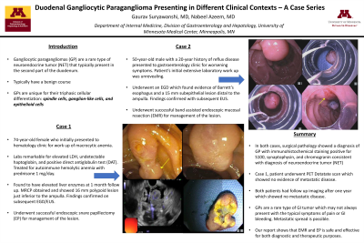
Introduction: Gangliocytic paragangliomas (GP) are a rare type of neuroendocrine tumor (NET) that typically present in the second part of the duodenum. While typically known to have a benign course, they are unique due to their triphasic cellular differentiation– spindle cells, ganglion-like cells, and epithelioid cells - thus require proper diagnosis to differentiate from other similar appearing duodenal tumors. We present two patients with GPs with different clinical contexts.
Case Description/Methods: Case 1: A 50-year-old-man with a 20-year history of reflux disease presented to gastroenterology clinic for worsening symptoms. The patient’s laboratory work up was otherwise unrevealing. Esophagogastroduodenoscopy (EGD) and EUS found evidence of Barrett’s esophagus and a 15 mm subepithelial lesion distal to the ampulla. Patient had a follow up EGD during which he underwent successful band-assisted endoscopic mucosal resection (EMR).
Case 2: A 74-year-old woman initially presented to hematology clinic for a new diagnosis of macrocytic anemia with a hemoglobin of 9.7 and MCV of 101. Extensive anemia work up was notable for normal folate and B12 but an elevated LDH and undetectable haptoglobin in the setting of a positive direct antiglobulin test (DAT). She was treated for autoimmune hemolytic anemia with prednisone 1 mg/day. During hematology follow up a few months later, the patient was found to have elevated liver enzymes which prompted an MR Abdomen (MRCP). MRCP showed a 16 mm polypoid lesion just inferior to the ampulla that was confirmed by subsequent endoscopic ultrasound (EUS). Patient underwent a successful endoscopic snare papillectomy (EP) for management of the lesion.
In both cases, surgical pathology showed diagnosis of GP with immunohistochemical staining positive for S100, synaptophysin, and chromogranin consistent with diagnosis of neuroendocrine tumor. In case 2, the patient underwent PET Dotatate scan which showed no evidence of metastatic disease. Both patients had follow up imaging after one year which showed no metastatic disease.
Discussion: GPs are a rare type of gastrointestinal tumor which may not always present with the common symptoms of abdominal pain and gastrointestinal bleeding. While typically benign, reports of metastatic spread are present. Our report shows that endoscopic management with either EMR or EP is safe and effective for both diagnostic and therapeutic purposes.

Disclosures:
Gaurav Suryawanshi, MD, Nabeel Azeem, MD. P4972 - Duodenal Gangliocytic Paragangliomas Presenting in Different Clinical Contexts- A Case Series, ACG 2024 Annual Scientific Meeting Abstracts. Philadelphia, PA: American College of Gastroenterology.
University of Minnesota Medical Center, Minneapolis, MN
Introduction: Gangliocytic paragangliomas (GP) are a rare type of neuroendocrine tumor (NET) that typically present in the second part of the duodenum. While typically known to have a benign course, they are unique due to their triphasic cellular differentiation– spindle cells, ganglion-like cells, and epithelioid cells - thus require proper diagnosis to differentiate from other similar appearing duodenal tumors. We present two patients with GPs with different clinical contexts.
Case Description/Methods: Case 1: A 50-year-old-man with a 20-year history of reflux disease presented to gastroenterology clinic for worsening symptoms. The patient’s laboratory work up was otherwise unrevealing. Esophagogastroduodenoscopy (EGD) and EUS found evidence of Barrett’s esophagus and a 15 mm subepithelial lesion distal to the ampulla. Patient had a follow up EGD during which he underwent successful band-assisted endoscopic mucosal resection (EMR).
Case 2: A 74-year-old woman initially presented to hematology clinic for a new diagnosis of macrocytic anemia with a hemoglobin of 9.7 and MCV of 101. Extensive anemia work up was notable for normal folate and B12 but an elevated LDH and undetectable haptoglobin in the setting of a positive direct antiglobulin test (DAT). She was treated for autoimmune hemolytic anemia with prednisone 1 mg/day. During hematology follow up a few months later, the patient was found to have elevated liver enzymes which prompted an MR Abdomen (MRCP). MRCP showed a 16 mm polypoid lesion just inferior to the ampulla that was confirmed by subsequent endoscopic ultrasound (EUS). Patient underwent a successful endoscopic snare papillectomy (EP) for management of the lesion.
In both cases, surgical pathology showed diagnosis of GP with immunohistochemical staining positive for S100, synaptophysin, and chromogranin consistent with diagnosis of neuroendocrine tumor. In case 2, the patient underwent PET Dotatate scan which showed no evidence of metastatic disease. Both patients had follow up imaging after one year which showed no metastatic disease.
Discussion: GPs are a rare type of gastrointestinal tumor which may not always present with the common symptoms of abdominal pain and gastrointestinal bleeding. While typically benign, reports of metastatic spread are present. Our report shows that endoscopic management with either EMR or EP is safe and effective for both diagnostic and therapeutic purposes.

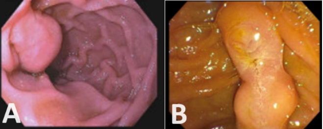
Figure: (A) Patient 1, 15 mm lesion distal to the ampulla. (B) Patient 2, 16 mm polypoid lesion just inferior to the ampulla.
Disclosures:
Gaurav Suryawanshi indicated no relevant financial relationships.
Nabeel Azeem: Boston Scientific – Consultant.
Gaurav Suryawanshi, MD, Nabeel Azeem, MD. P4972 - Duodenal Gangliocytic Paragangliomas Presenting in Different Clinical Contexts- A Case Series, ACG 2024 Annual Scientific Meeting Abstracts. Philadelphia, PA: American College of Gastroenterology.
