Tuesday Poster Session
Category: Small Intestine
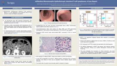
P4987 - Infiltrative Monomorphic Epitheliotropic Intestinal T-Cell Lymphoma: A Case Report
Tuesday, October 29, 2024
10:30 AM - 4:00 PM ET
Location: Exhibit Hall E

Has Audio

Nabil El Hage Chehade, MD
Scripps Clinic
San Diego, CA
Presenting Author(s)
Nabil El Hage Chehade, MD1, Vandan Patel, MD1, Olivia Lanser, MD2, Pranav Gandhi, MD3, Matthew Skinner, MD4, Gauree Konijeti, MD, MPH3
1Scripps Clinic, San Diego, CA; 2Scripps Green Hospital, La Jolla, CA; 3Scripps Clinic, La Jolla, CA; 4Scripps Clinic & Scripps Research Institute, San Diego, CA
Introduction: Monomorphic epitheliotropic intestinal T-cell lymphoma (MEITL) is a rare aggressive T-cell lymphoma more common in Asian and Hispanic populations. Review of the literature suggests most cases of MEITL manifest as small bowel tumors and present with bowel obstruction or perforation. Here, we present a case of a Caucasian patient with subacute diarrhea who was diagnosed with malignant MEITL with ileocolonic and bone marrow involvement.
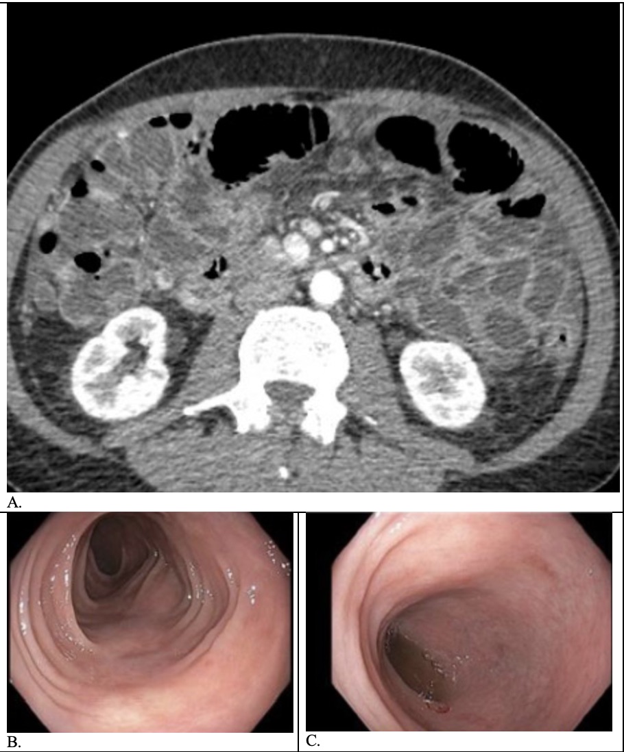
Case Description/Methods: An 80-year-old man with hypertension was hospitalized with 3 weeks of severe subacute watery diarrhea, abdominal discomfort, and chills. Labs showed only mild hyponatremia, with negative celiac panel. Stool studies were negative for infection, with normal elastase and elevated calprotectin at 1200 ug/g. CT abdomen/pelvis with IV contrast demonstrated moderate-to-severe ileal thickening and enhancement without small bowel obstruction. Colonoscopy revealed normal colon but edema, friable mucosa and villous blunting in the terminal ileum. Histopathology from the colon and ileum demonstrated dense, diffuse monomorphic atypical lymphoid cells without necrosis. Immunohistochemical stains were positive for CD8, CD56, and T-cell receptor gamma/beta rearrangement. There was dim nuclear expression of TP53, and MYC was negative. Subsequent bone marrow biopsy demonstrated MEITL composing 10-15% of marrow cellularity. With diagnosis of malignant MEITL, patient was initiated on CHOP chemotherapy. Course was complicated by recurrent C. difficile infection, neutropenic fever, pulmonary embolism, and failure to thrive due to persistent severe diarrhea despite various measures. After 2 months, patient was eventually transitioned to hospice.
Discussion: MEITL is an aggressive malignant disease often resistant to conventional chemotherapy. MYC expression and TP53 mutation are associated with worse prognosis. Our patient’s presentation of MEITL was atypical, with subacute diarrhea, subtle endoscopic findings but marked radiographic transmural ileal thickening. This case highlights the importance of timely evaluation and close multidisciplinary collaboration to facilitate diagnosis and management. Further research into more effective therapies for MEITL is warranted.

Disclosures:
Nabil El Hage Chehade, MD1, Vandan Patel, MD1, Olivia Lanser, MD2, Pranav Gandhi, MD3, Matthew Skinner, MD4, Gauree Konijeti, MD, MPH3. P4987 - Infiltrative Monomorphic Epitheliotropic Intestinal T-Cell Lymphoma: A Case Report, ACG 2024 Annual Scientific Meeting Abstracts. Philadelphia, PA: American College of Gastroenterology.
1Scripps Clinic, San Diego, CA; 2Scripps Green Hospital, La Jolla, CA; 3Scripps Clinic, La Jolla, CA; 4Scripps Clinic & Scripps Research Institute, San Diego, CA
Introduction: Monomorphic epitheliotropic intestinal T-cell lymphoma (MEITL) is a rare aggressive T-cell lymphoma more common in Asian and Hispanic populations. Review of the literature suggests most cases of MEITL manifest as small bowel tumors and present with bowel obstruction or perforation. Here, we present a case of a Caucasian patient with subacute diarrhea who was diagnosed with malignant MEITL with ileocolonic and bone marrow involvement.
Case Description/Methods: An 80-year-old man with hypertension was hospitalized with 3 weeks of severe subacute watery diarrhea, abdominal discomfort, and chills. Labs showed only mild hyponatremia, with negative celiac panel. Stool studies were negative for infection, with normal elastase and elevated calprotectin at 1200 ug/g. CT abdomen/pelvis with IV contrast demonstrated moderate-to-severe ileal thickening and enhancement without small bowel obstruction. Colonoscopy revealed normal colon but edema, friable mucosa and villous blunting in the terminal ileum. Histopathology from the colon and ileum demonstrated dense, diffuse monomorphic atypical lymphoid cells without necrosis. Immunohistochemical stains were positive for CD8, CD56, and T-cell receptor gamma/beta rearrangement. There was dim nuclear expression of TP53, and MYC was negative. Subsequent bone marrow biopsy demonstrated MEITL composing 10-15% of marrow cellularity. With diagnosis of malignant MEITL, patient was initiated on CHOP chemotherapy. Course was complicated by recurrent C. difficile infection, neutropenic fever, pulmonary embolism, and failure to thrive due to persistent severe diarrhea despite various measures. After 2 months, patient was eventually transitioned to hospice.
Discussion: MEITL is an aggressive malignant disease often resistant to conventional chemotherapy. MYC expression and TP53 mutation are associated with worse prognosis. Our patient’s presentation of MEITL was atypical, with subacute diarrhea, subtle endoscopic findings but marked radiographic transmural ileal thickening. This case highlights the importance of timely evaluation and close multidisciplinary collaboration to facilitate diagnosis and management. Further research into more effective therapies for MEITL is warranted.

Figure: (A) CT abdomen and pelvis with IV contrast demonstrating moderate-to-severe ileal thickening and enhancement without small bowel obstruction; (B-C) Colonoscopy demonstrating normal colon but edema, friable mucosa and villous blunting in the terminal ileum
Disclosures:
Nabil El Hage Chehade indicated no relevant financial relationships.
Vandan Patel indicated no relevant financial relationships.
Olivia Lanser indicated no relevant financial relationships.
Pranav Gandhi indicated no relevant financial relationships.
Matthew Skinner indicated no relevant financial relationships.
Gauree Konijeti: Abbvie – Advisory Committee/Board Member, Consultant. Lilly – Consultant, Speakers Bureau. Pfizer – Advisory Committee/Board Member. Takeda – Speakers Bureau.
Nabil El Hage Chehade, MD1, Vandan Patel, MD1, Olivia Lanser, MD2, Pranav Gandhi, MD3, Matthew Skinner, MD4, Gauree Konijeti, MD, MPH3. P4987 - Infiltrative Monomorphic Epitheliotropic Intestinal T-Cell Lymphoma: A Case Report, ACG 2024 Annual Scientific Meeting Abstracts. Philadelphia, PA: American College of Gastroenterology.
