Tuesday Poster Session
Category: Small Intestine
P5000 - Superior Mesenteric Vein Syndrome, A Case of Small Bowel Obstruction
Tuesday, October 29, 2024
10:30 AM - 4:00 PM ET
Location: Exhibit Hall E

Has Audio
- PP
Parth Patel, MD
University of Texas Health San Antonio
San Antonio, TX
Presenting Author(s)
Parth Patel, MD1, Adam Vinall, MD2, Arham Siddiqui, MD1, Saatchi Kuwelker, MD2, Juan Echavarria, MD1
1University of Texas Health San Antonio, San Antonio, TX; 2University of Texas Health Science Center, San Antonio, TX
Introduction: Superior Mesenteric Artery (SMA) Syndrome is a rare condition where the third portion of the duodenum becomes compressed between the SMA and the aorta. Symptomology is vague and can be mimicked by the even lesser-known Superior Mesenteric Vein (SMV) Syndrome where the third portion of the duodenum is compressed by an aberrant SMV. In this report, we present a unique case of SMV syndrome.
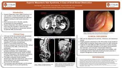
Case Description/Methods: A 60-year-old female presents with abdominal pain, nausea, and vomiting. History is pertinent for prior small bowel volvulus complicated by gangrenous small bowel infarction resulting in resection of 10 inches of small bowel. An upper GI series revealed partial obstruction at the level of the third portion of the duodenum concerning for SMA syndrome. A CT abdomen/pelvis with IV contrast showed diffuse fluid-filled dilatation of the proximal duodenal segment extending into the third portion of the duodenum prior to crossing the SMV. The SMA was not involved in this process as confirmed by two separate radiologists. (Figure 1) Despite nasogastric tube decompression, symptoms persisted with notable bilious output. Patient was taken for esophagogastroduodenoscopy which showed extrinsic compression of the 3rd portion of the duodenum with a visible pulsatile mass causing proximal dilation. (Figure 2) The patient subsequently underwent robotic-assisted laparoscopic diagnostic laparoscopy, lysis of small bowel adhesions, and duodenojejunostomy proximal to the location of the obstruction. The procedure was tolerated well, and the patient was discharged thirty-six hours post-operatively.
Discussion: Small bowel obstruction (SBO) can be broken down into 3 general categories; extrinsic, intramural, and intraluminal. In this case, we present a less commonly seen source of extrinsic SBO with an initial presentation concerning for SMA syndrome. However, imaging was significant for compression of the third portion of the duodenum without involvement of the SMA raising concern for SMV syndrome. Treatment of both SMA and SMV syndrome are identical with early focus on conservative therapies such as gastric decompression, fluid restriction and correction of electrolyte abnormalities. If conservative management fails, several surgical options are available, with duodenojejunostomy historically providing the best results.

Disclosures:
Parth Patel, MD1, Adam Vinall, MD2, Arham Siddiqui, MD1, Saatchi Kuwelker, MD2, Juan Echavarria, MD1. P5000 - Superior Mesenteric Vein Syndrome, A Case of Small Bowel Obstruction, ACG 2024 Annual Scientific Meeting Abstracts. Philadelphia, PA: American College of Gastroenterology.
1University of Texas Health San Antonio, San Antonio, TX; 2University of Texas Health Science Center, San Antonio, TX
Introduction: Superior Mesenteric Artery (SMA) Syndrome is a rare condition where the third portion of the duodenum becomes compressed between the SMA and the aorta. Symptomology is vague and can be mimicked by the even lesser-known Superior Mesenteric Vein (SMV) Syndrome where the third portion of the duodenum is compressed by an aberrant SMV. In this report, we present a unique case of SMV syndrome.
Case Description/Methods: A 60-year-old female presents with abdominal pain, nausea, and vomiting. History is pertinent for prior small bowel volvulus complicated by gangrenous small bowel infarction resulting in resection of 10 inches of small bowel. An upper GI series revealed partial obstruction at the level of the third portion of the duodenum concerning for SMA syndrome. A CT abdomen/pelvis with IV contrast showed diffuse fluid-filled dilatation of the proximal duodenal segment extending into the third portion of the duodenum prior to crossing the SMV. The SMA was not involved in this process as confirmed by two separate radiologists. (Figure 1) Despite nasogastric tube decompression, symptoms persisted with notable bilious output. Patient was taken for esophagogastroduodenoscopy which showed extrinsic compression of the 3rd portion of the duodenum with a visible pulsatile mass causing proximal dilation. (Figure 2) The patient subsequently underwent robotic-assisted laparoscopic diagnostic laparoscopy, lysis of small bowel adhesions, and duodenojejunostomy proximal to the location of the obstruction. The procedure was tolerated well, and the patient was discharged thirty-six hours post-operatively.
Discussion: Small bowel obstruction (SBO) can be broken down into 3 general categories; extrinsic, intramural, and intraluminal. In this case, we present a less commonly seen source of extrinsic SBO with an initial presentation concerning for SMA syndrome. However, imaging was significant for compression of the third portion of the duodenum without involvement of the SMA raising concern for SMV syndrome. Treatment of both SMA and SMV syndrome are identical with early focus on conservative therapies such as gastric decompression, fluid restriction and correction of electrolyte abnormalities. If conservative management fails, several surgical options are available, with duodenojejunostomy historically providing the best results.

Figure: Figure 1: 3rd portion of duodenum compressed by Aorta and SMV with distal obstruction
Figure 2: Endoscopic image of 3rd portion of duodenum with visible enlarged folds compressing lumen
Figure 2: Endoscopic image of 3rd portion of duodenum with visible enlarged folds compressing lumen
Disclosures:
Parth Patel indicated no relevant financial relationships.
Adam Vinall indicated no relevant financial relationships.
Arham Siddiqui indicated no relevant financial relationships.
Saatchi Kuwelker indicated no relevant financial relationships.
Juan Echavarria indicated no relevant financial relationships.
Parth Patel, MD1, Adam Vinall, MD2, Arham Siddiqui, MD1, Saatchi Kuwelker, MD2, Juan Echavarria, MD1. P5000 - Superior Mesenteric Vein Syndrome, A Case of Small Bowel Obstruction, ACG 2024 Annual Scientific Meeting Abstracts. Philadelphia, PA: American College of Gastroenterology.
