Tuesday Poster Session
Category: Small Intestine
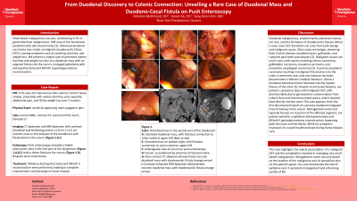
P5024 - From Duodenal Discovery to Colonic Connection: Unveiling a Rare Case of Duodenal Mass and Duodeno-Cecal Fistula on Push Enteroscopy
Tuesday, October 29, 2024
10:30 AM - 4:00 PM ET
Location: Exhibit Hall E

Has Audio

Abrahim Mahmood, DO
New York-Presbyterian/Queens
Queens, NY
Presenting Author(s)
Abrahim Mahmood, DO1, Hasan Ali, DO2, Sang Hoon Kim, MD2
1New York-Presbyterian/Queens, Queens, NY; 2New York-Presbyterian/Queens, Flushing, NY
Introduction: Small bowel malignancies are rare, constituting 3-5% of gastrointestinal malignancies. Half arise in the duodenum, predominantly adenocarcinomas. Advanced duodenal carcinoma may create a malignant duodenocolic fistula (DCF), causing symptoms such as vomiting, diarrhea, and weight loss. We present a unique case of persistent watery diarrhea and weight loss due to a duodenal mass with an adjacent fistula into the cecum, managed palliatively with distal gastrectomy and billroth II gastrojejunostomy reconstruction.
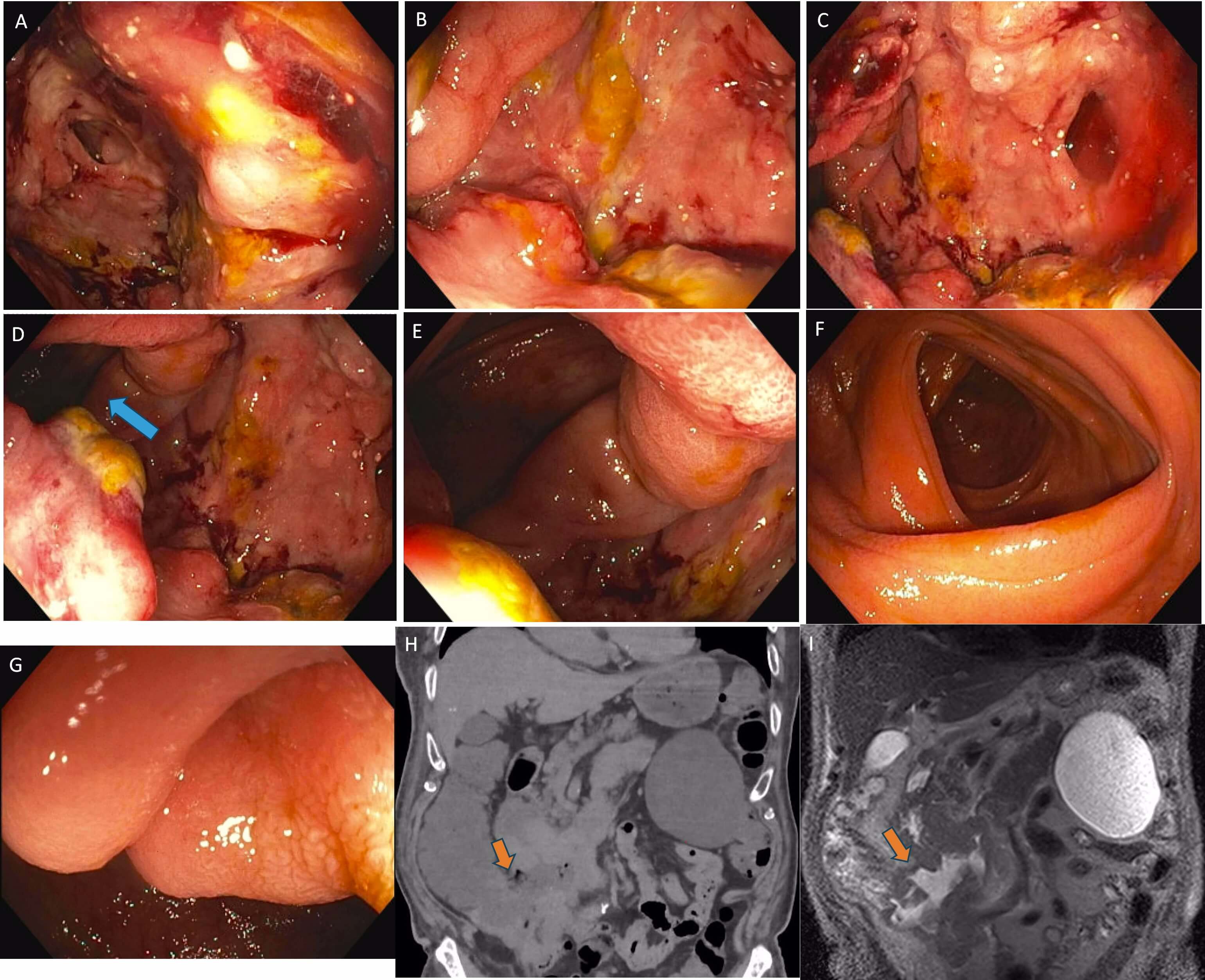
Case Description/Methods: A 91-year-old male Korean War veteran, former heavy smoker, presented with watery diarrhea, abdominal pain, and 40 lbs weight loss over 2 months. Physical exam revealed epigastric pain and cachexia. Imaging showed duodenal wall thickening and an 11.8 cm x 11.6 cm necrotic mass in the 2nd part of the duodenum with fistulization to the cecum (Figure H,I). Push enteroscopy revealed a friable obstructive mass in the 2nd part of the duodenum (Figure A,B,C) with a direct fistula to the cecum (Figure D). Biopsies were inconclusive. Palliative distal gastrectomy and billroth II reconstruction were performed, leading to symptom improvement and discharge to home hospice.
Discussion: Duodenal malignancies, predominantly adenocarcinomas, are rare, with DCF formation even rarer. Our patient's symptoms align with malignant DCF presentation, likely due to gastroenteric contamination with colonic flora and mechanical obstruction shunting food contents directly into the colon. We believe our case is the first documented report of a primary duodenal malignant mass fistulizing to the cecum. Management varies; our patient opted for palliative distal gastrectomy and billroth II gastrojejunostomy reconstruction, bypassing the mass and fistula. While symptoms improved, overall health declined during home hospice. This case highlights the typical presentation of a malignant DCF and the complexity of managing rare small bowel malignancies. It emphasizes palliative care’s role in symptom management and quality of life.

Disclosures:
Abrahim Mahmood, DO1, Hasan Ali, DO2, Sang Hoon Kim, MD2. P5024 - From Duodenal Discovery to Colonic Connection: Unveiling a Rare Case of Duodenal Mass and Duodeno-Cecal Fistula on Push Enteroscopy, ACG 2024 Annual Scientific Meeting Abstracts. Philadelphia, PA: American College of Gastroenterology.
1New York-Presbyterian/Queens, Queens, NY; 2New York-Presbyterian/Queens, Flushing, NY
Introduction: Small bowel malignancies are rare, constituting 3-5% of gastrointestinal malignancies. Half arise in the duodenum, predominantly adenocarcinomas. Advanced duodenal carcinoma may create a malignant duodenocolic fistula (DCF), causing symptoms such as vomiting, diarrhea, and weight loss. We present a unique case of persistent watery diarrhea and weight loss due to a duodenal mass with an adjacent fistula into the cecum, managed palliatively with distal gastrectomy and billroth II gastrojejunostomy reconstruction.
Case Description/Methods: A 91-year-old male Korean War veteran, former heavy smoker, presented with watery diarrhea, abdominal pain, and 40 lbs weight loss over 2 months. Physical exam revealed epigastric pain and cachexia. Imaging showed duodenal wall thickening and an 11.8 cm x 11.6 cm necrotic mass in the 2nd part of the duodenum with fistulization to the cecum (Figure H,I). Push enteroscopy revealed a friable obstructive mass in the 2nd part of the duodenum (Figure A,B,C) with a direct fistula to the cecum (Figure D). Biopsies were inconclusive. Palliative distal gastrectomy and billroth II reconstruction were performed, leading to symptom improvement and discharge to home hospice.
Discussion: Duodenal malignancies, predominantly adenocarcinomas, are rare, with DCF formation even rarer. Our patient's symptoms align with malignant DCF presentation, likely due to gastroenteric contamination with colonic flora and mechanical obstruction shunting food contents directly into the colon. We believe our case is the first documented report of a primary duodenal malignant mass fistulizing to the cecum. Management varies; our patient opted for palliative distal gastrectomy and billroth II gastrojejunostomy reconstruction, bypassing the mass and fistula. While symptoms improved, overall health declined during home hospice. This case highlights the typical presentation of a malignant DCF and the complexity of managing rare small bowel malignancies. It emphasizes palliative care’s role in symptom management and quality of life.

Figure: A,B,C: Ulcerated mass in the second part of the duodenum D: Ulcerated duodenal mass, with fistulous connection to colon noted at upper-left (blue arrow) E: Ulcerated mass on bottom right, with fistulous connection to colon noted on upper-left F: Anterograde view of cecum on push enteroscopy G: Cecum, as evidenced by presence of ileocecal valve H: Non-contrast CT image demonstrates necrotic duodenal mass with duodenocolic fistula (orange arrow) I: Contrast-enhanced MRI Abdomen demonstrates necrotic duodenal mass with duodenocolic fistula (orange arrow)
Disclosures:
Abrahim Mahmood indicated no relevant financial relationships.
Hasan Ali indicated no relevant financial relationships.
Sang Hoon Kim indicated no relevant financial relationships.
Abrahim Mahmood, DO1, Hasan Ali, DO2, Sang Hoon Kim, MD2. P5024 - From Duodenal Discovery to Colonic Connection: Unveiling a Rare Case of Duodenal Mass and Duodeno-Cecal Fistula on Push Enteroscopy, ACG 2024 Annual Scientific Meeting Abstracts. Philadelphia, PA: American College of Gastroenterology.
