Tuesday Poster Session
Category: Small Intestine
P5035 - Secondary Intestinal Lymphangiectasia Due to Waldenström Macroglobulinemia
Tuesday, October 29, 2024
10:30 AM - 4:00 PM ET
Location: Exhibit Hall E

Has Audio

Samuel Y. Jo, MD
Robert Wood Johnson Medical School, Rutgers University
New Brunswick, NJ
Presenting Author(s)
Samuel Y. Jo, MD, Isago Jerrett, MD, Sarag Boukhar, MD, Anish V. Patel, MD
Robert Wood Johnson Medical School, Rutgers University, New Brunswick, NJ
Introduction: Intestinal lymphangiectasia is a condition characterized by pathologic dilation of lacteals causing drainage of lymphatic fluid into the small bowel. This can cause vague gastro-intestinal symptoms as well as systemic symptoms not limited to diarrhea, abdominal pain, edema and fatigue. Intestinal lymphangiectasia can occur as a primary disorder from congenital malformation of the lymphatic channel or secondary due to lymphatic obstruction from conditions such as right sided heart failure, cirrhosis, retroperitoneal fibrosis, and neoplasia involving the lymph nodes. We present a case of intestinal lymphangiectasia in a patient newly diagnosed with Waldenstrom’s Macroglobulinemia.
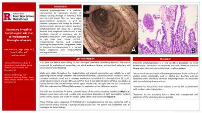
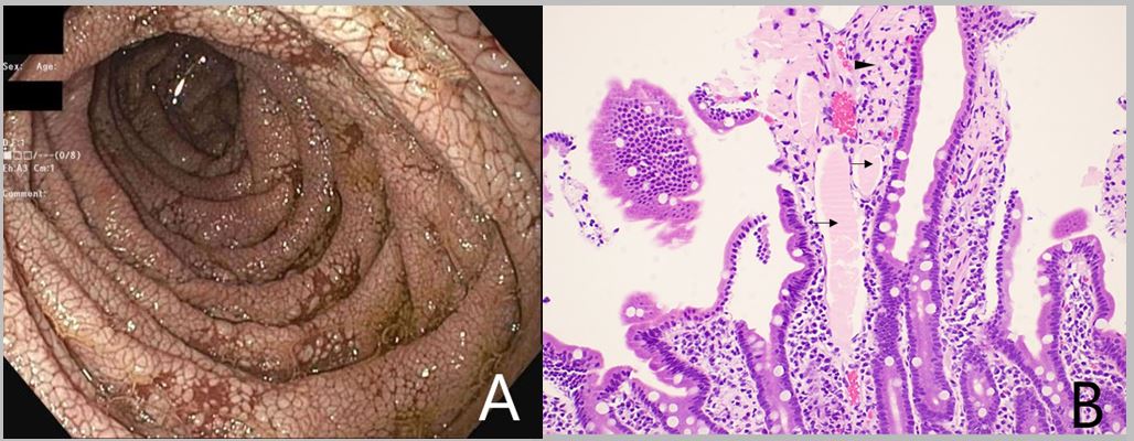
Case Description/Methods: A 61 year old female with history of anti-cardiolipin syndrome, pulmonary embolus, and MGUS presented for evaluation of worsening generalized weakness, fatigue, unintentional weightless and small volume rectal bleeding. Vitals were stable and physical examination was notable for a frail appearing female, benign abdomen and external hemorrhoids. Laboratory results revealed a normal complete metabolic panel, and a complete blood count remarkable for a hemoglobin of 11.2 g/dL, white blood cell count of 3.59 x 10^3/uL, MCV of 92.5 fL and platelets 220 x 10^3/uL. Iron studies a normal ferritin of 98, borderline low Iron 44 ug/dl, normal TIBC 287 ug/dl and low iron saturation of 15%. She underwent an EGD and Colonoscopy for evaluation of iron deficiency anemia. The EGD was remarkable for white colored mucosa of the duodenum (Figure A). Biopsies were taken and resulted with amorphous deposition of light eosinophilic material within the lamina propria and inside the dilated lacteal of the small bowel villi (Figure B). These findings were suggestive of Waldenstrom’s Macroglobulinemia and later confirmed with a bone marrow biopsy showing > 10% lymphoplasmacytic cell.
Discussion: Intestinal lymphangiectasia is a rare condition diagnosed via small bowel biopsy. The lesions can be patchy in nature, therefore a normal biopsy alone does not rule out intestinal lymphangiectasia. Symptoms of primary intestinal lymphangiectasia are similar to those of protein losing enteropathy such as edema and diarrhea, whereas symptoms from secondary intestinal lymphangiectasia are associated with the underlying disease process. Treatment for the primary form includes a low fat diet supplemented with medium-chain triglycerides. Treatment for the secondary form involves resolving the underlying disease process.

Disclosures:
Samuel Y. Jo, MD, Isago Jerrett, MD, Sarag Boukhar, MD, Anish V. Patel, MD. P5035 - Secondary Intestinal Lymphangiectasia Due to Waldenström Macroglobulinemia, ACG 2024 Annual Scientific Meeting Abstracts. Philadelphia, PA: American College of Gastroenterology.
Robert Wood Johnson Medical School, Rutgers University, New Brunswick, NJ
Introduction: Intestinal lymphangiectasia is a condition characterized by pathologic dilation of lacteals causing drainage of lymphatic fluid into the small bowel. This can cause vague gastro-intestinal symptoms as well as systemic symptoms not limited to diarrhea, abdominal pain, edema and fatigue. Intestinal lymphangiectasia can occur as a primary disorder from congenital malformation of the lymphatic channel or secondary due to lymphatic obstruction from conditions such as right sided heart failure, cirrhosis, retroperitoneal fibrosis, and neoplasia involving the lymph nodes. We present a case of intestinal lymphangiectasia in a patient newly diagnosed with Waldenstrom’s Macroglobulinemia.
Case Description/Methods: A 61 year old female with history of anti-cardiolipin syndrome, pulmonary embolus, and MGUS presented for evaluation of worsening generalized weakness, fatigue, unintentional weightless and small volume rectal bleeding. Vitals were stable and physical examination was notable for a frail appearing female, benign abdomen and external hemorrhoids. Laboratory results revealed a normal complete metabolic panel, and a complete blood count remarkable for a hemoglobin of 11.2 g/dL, white blood cell count of 3.59 x 10^3/uL, MCV of 92.5 fL and platelets 220 x 10^3/uL. Iron studies a normal ferritin of 98, borderline low Iron 44 ug/dl, normal TIBC 287 ug/dl and low iron saturation of 15%. She underwent an EGD and Colonoscopy for evaluation of iron deficiency anemia. The EGD was remarkable for white colored mucosa of the duodenum (Figure A). Biopsies were taken and resulted with amorphous deposition of light eosinophilic material within the lamina propria and inside the dilated lacteal of the small bowel villi (Figure B). These findings were suggestive of Waldenstrom’s Macroglobulinemia and later confirmed with a bone marrow biopsy showing > 10% lymphoplasmacytic cell.
Discussion: Intestinal lymphangiectasia is a rare condition diagnosed via small bowel biopsy. The lesions can be patchy in nature, therefore a normal biopsy alone does not rule out intestinal lymphangiectasia. Symptoms of primary intestinal lymphangiectasia are similar to those of protein losing enteropathy such as edema and diarrhea, whereas symptoms from secondary intestinal lymphangiectasia are associated with the underlying disease process. Treatment for the primary form includes a low fat diet supplemented with medium-chain triglycerides. Treatment for the secondary form involves resolving the underlying disease process.

Figure: Figure A: Endoscopic view of small bowel.
Figure B: High power view of small bowel villi with findings of deposits of amorphous, light eosinophilic material within lamina propria (arrowhead) and within dilated lacteals (arrow).
Figure B: High power view of small bowel villi with findings of deposits of amorphous, light eosinophilic material within lamina propria (arrowhead) and within dilated lacteals (arrow).
Disclosures:
Samuel Jo indicated no relevant financial relationships.
Isago Jerrett indicated no relevant financial relationships.
Sarag Boukhar indicated no relevant financial relationships.
Anish Patel indicated no relevant financial relationships.
Samuel Y. Jo, MD, Isago Jerrett, MD, Sarag Boukhar, MD, Anish V. Patel, MD. P5035 - Secondary Intestinal Lymphangiectasia Due to Waldenström Macroglobulinemia, ACG 2024 Annual Scientific Meeting Abstracts. Philadelphia, PA: American College of Gastroenterology.
