Tuesday Poster Session
Category: Small Intestine
P5037 - A Hidden Carcinoid Tumor Debuting With Persistent, Overt Small Bowel Bleeding: A Three Capsule Endoscopy Case
Tuesday, October 29, 2024
10:30 AM - 4:00 PM ET
Location: Exhibit Hall E

Has Audio
- JF
Juan G. Feliciano Figueroa, MD
VA Caribbean Healthcare System
San Juan, PR
Presenting Author(s)
Juan G. Feliciano Figueroa, MD, Carla Cepero Jimenez, MD, Carolina Diaz Loza, MD, Jose Martin-Ortiz, MD, FACG
VA Caribbean Healthcare System, San Juan, Puerto Rico
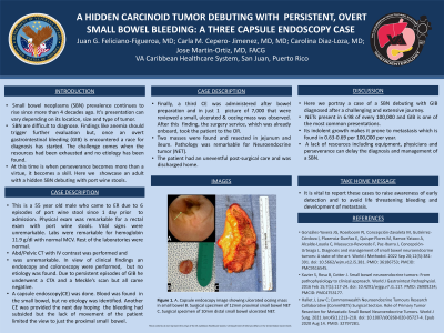
Introduction: Small bowel neoplasms (SBN) prevalence continues to rise since more than 4 decades ago. It’s presentation can vary depending on its location, size and type of tumor. The low conspicuity of SBN contributes to a late diagnosis. Findings like anemia should trigger further evaluation but, once an overt gastrointestinal bleeding (GIB) is encountered a race for diagnosis has started. The challenge comes when the resources had been exhausted and no etiology has been found. At this time is when perseverance becomes more than a virtue, it becomes a skill. Here we showcase an adult with a hidden SBN debuting with port wine stools.
Case Description/Methods: This is a 55 year old male who came to ER due to 6 episodes of port wine stool since 1 day prior to admission. Physical exam was remarkable for a rectal exam with port wine stools. Rest of the exam and vital signs were unremarkable. Labs were remarkable for hemoglobin 11.9 with normal MCV. Rest of the laboratories were normal. Abd/Pelvic CT with IV contrast was performed and was unremarkable. In view of clinical findings an endoscopy and colonoscopy were performed, but no etiology was found. Due to persistent episodes of GIB he underwent a CTA, angiography and a Meckle’s scan but all came negative. A capsule endoscopy(CE) was done. Blood was found in the small bowel, but no etiology was identified. Another CE was provided the next day hoping the bleeding had subsided but the lack of movement of the patient limited the view to just the proximal small bowel. Finally, a third CE was administered after bowel preparation and in just 1 picture of 7,000 that were reviewed a small, ulcerated & oozing mass was observed. After this finding, the surgery service, which was already onboard, took the patient to the OR. Two masses were found and resected in jejunum and ileum. Pathology was remarkable for Neuroendocrine tumor (NET). The patient had an uneventful post-surgical care and was discharged home.
Discussion: Here we portray a case of a SBN debuting with GIB diagnosed after a challenging and extensive journey. NETs present in 6.98 of every 100,000 and GIB is one of the most common presentations. Its indolent growth makes it prone to metastasis which is found in 0.63-0.69 per 100,000 per year. A lack of resources including equipment, physicians and perseverance can delay the diagnosis and management of a SBN. It is vital to report these cases to raise awareness of early detection of NETs to avoid life threatening bleeding and development of metastasis.

Disclosures:
Juan G. Feliciano Figueroa, MD, Carla Cepero Jimenez, MD, Carolina Diaz Loza, MD, Jose Martin-Ortiz, MD, FACG. P5037 - A Hidden Carcinoid Tumor Debuting With Persistent, Overt Small Bowel Bleeding: A Three Capsule Endoscopy Case, ACG 2024 Annual Scientific Meeting Abstracts. Philadelphia, PA: American College of Gastroenterology.
VA Caribbean Healthcare System, San Juan, Puerto Rico
Introduction: Small bowel neoplasms (SBN) prevalence continues to rise since more than 4 decades ago. It’s presentation can vary depending on its location, size and type of tumor. The low conspicuity of SBN contributes to a late diagnosis. Findings like anemia should trigger further evaluation but, once an overt gastrointestinal bleeding (GIB) is encountered a race for diagnosis has started. The challenge comes when the resources had been exhausted and no etiology has been found. At this time is when perseverance becomes more than a virtue, it becomes a skill. Here we showcase an adult with a hidden SBN debuting with port wine stools.
Case Description/Methods: This is a 55 year old male who came to ER due to 6 episodes of port wine stool since 1 day prior to admission. Physical exam was remarkable for a rectal exam with port wine stools. Rest of the exam and vital signs were unremarkable. Labs were remarkable for hemoglobin 11.9 with normal MCV. Rest of the laboratories were normal. Abd/Pelvic CT with IV contrast was performed and was unremarkable. In view of clinical findings an endoscopy and colonoscopy were performed, but no etiology was found. Due to persistent episodes of GIB he underwent a CTA, angiography and a Meckle’s scan but all came negative. A capsule endoscopy(CE) was done. Blood was found in the small bowel, but no etiology was identified. Another CE was provided the next day hoping the bleeding had subsided but the lack of movement of the patient limited the view to just the proximal small bowel. Finally, a third CE was administered after bowel preparation and in just 1 picture of 7,000 that were reviewed a small, ulcerated & oozing mass was observed. After this finding, the surgery service, which was already onboard, took the patient to the OR. Two masses were found and resected in jejunum and ileum. Pathology was remarkable for Neuroendocrine tumor (NET). The patient had an uneventful post-surgical care and was discharged home.
Discussion: Here we portray a case of a SBN debuting with GIB diagnosed after a challenging and extensive journey. NETs present in 6.98 of every 100,000 and GIB is one of the most common presentations. Its indolent growth makes it prone to metastasis which is found in 0.63-0.69 per 100,000 per year. A lack of resources including equipment, physicians and perseverance can delay the diagnosis and management of a SBN. It is vital to report these cases to raise awareness of early detection of NETs to avoid life threatening bleeding and development of metastasis.

Figure: A. Capsule endoscopy image showing ulcerated oozing mass in small bowel B. Surgical specimen of 12mm proximal small bowel NET C. Surgical specimen of 10mm distal small bowel ulcerated NET.
Disclosures:
Juan Feliciano Figueroa indicated no relevant financial relationships.
Carla Cepero Jimenez indicated no relevant financial relationships.
Carolina Diaz Loza indicated no relevant financial relationships.
Jose Martin-Ortiz indicated no relevant financial relationships.
Juan G. Feliciano Figueroa, MD, Carla Cepero Jimenez, MD, Carolina Diaz Loza, MD, Jose Martin-Ortiz, MD, FACG. P5037 - A Hidden Carcinoid Tumor Debuting With Persistent, Overt Small Bowel Bleeding: A Three Capsule Endoscopy Case, ACG 2024 Annual Scientific Meeting Abstracts. Philadelphia, PA: American College of Gastroenterology.
