Tuesday Poster Session
Category: Stomach
P5110 - Chronic Mesenteric Ischemia Presenting With Multiple Gastric Ulcers: A Unique Case
Tuesday, October 29, 2024
10:30 AM - 4:00 PM ET
Location: Exhibit Hall E

Has Audio

Jyotroop Kaur, MBBS
University of South Dakota Sanford School of Medicine
Sioux Falls, SD
Presenting Author(s)
Jyotroop Kaur, MBBS, Kennedy Forest, MD, Rafia Irfan Waheed, MBBS, Jessica Pierobon, MD
University of South Dakota Sanford School of Medicine, Sioux Falls, SD
Introduction: Atherosclerotic disease affecting the mesenteric vasculature can result in chronic ischemia throughout different regions of the gastrointestinal system, although it rarely impacts the stomach. We present an unusual presentation of chronic mesenteric ischemia (CMI) with extensive gastric ulceration, a large cecal ulcer, and ischemic liver lesions.
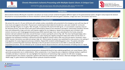
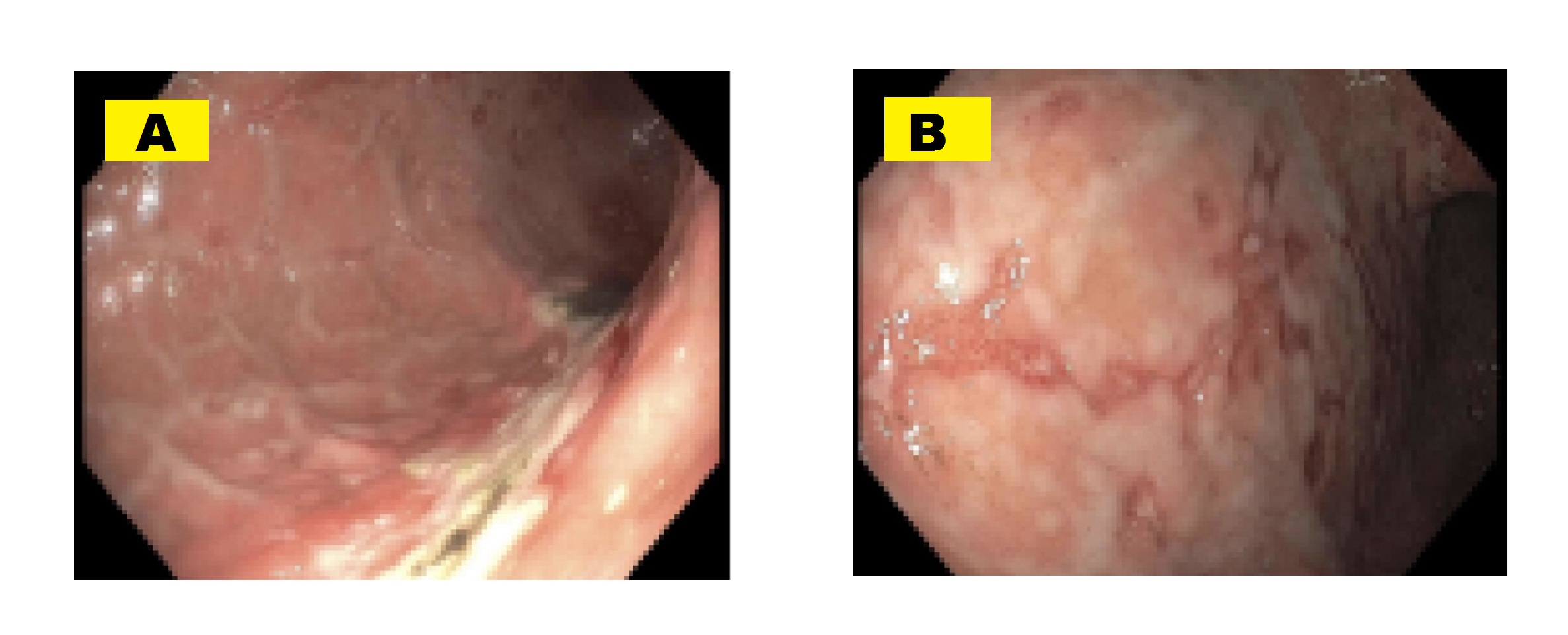
Case Description/Methods: We present the case of a 74-year-old female with multiple cardiac risk factors who presented to the ER with 2 days of loose stools and epigastric discomfort. Physical exam revealed generalized abdominal tenderness. Labs were notable for normocytic anemia with Hgb 9.8 g/dL, Fe 14 ug/dL, TIBC 156 ug/dL, Fe saturation 9% and Ferritin 829 ng/mL. CT scan showed a suspicious mass at the ileocecal junction, new liver lesions with severe atheromatous disease of the mesenteric vasculature. Colonoscopy revealed a necrotic mass and subsequently a large cecal ulcer with biopsies negative for malignancy. Hepatic tissue biopsies were consistent with ischemic necrosis as well. EGD reported large, linear ulcers extending from the fundus along the posterior wall and scattered small ulcers around the lesser curvature sparing the antrum (Fig.1). The ulcers had heaped edges and a clean base with biopsies showing reactive gastropathy. H. pylori testing was negative and gastrin levels were within normal limits. CT angiogram and subsequent mesenteric ultrasound confirmed significant stenosis within celiac axis and superior mesenteric artery (SMA). Vascular intervention was deferred owing to high risk. Unfortunately, the patient was readmitted for acute anemia in the setting of anticoagulation for newly diagnosed pulmonary embolism. Repeat EGD revealed nonbleeding gastric ulcers. The patient ultimately underwent successful IR-guided shockwave lithotripsy and stent reconstruction to the SMA in a technically difficult and complicated procedure. She has been recovering well for 2 months.
Discussion: This is a case of CMI with an atypical presentation subsequently found to have multi-vessel atherosclerosis. CMI is generally asymptomatic due to collateralization. When symptomatic, it can pose a diagnostic challenge leading to significant morbidity and delay in treatment. Our case highlights the importance of high suspicion for CMI when managing unexplained gastric ulcer disease, especially in patients with extensive risk factors. Alternate etiologies like NSAID usage, H. pylori infection and Zollinger-Ellison syndrome should be excluded.

Disclosures:
Jyotroop Kaur, MBBS, Kennedy Forest, MD, Rafia Irfan Waheed, MBBS, Jessica Pierobon, MD. P5110 - Chronic Mesenteric Ischemia Presenting With Multiple Gastric Ulcers: A Unique Case, ACG 2024 Annual Scientific Meeting Abstracts. Philadelphia, PA: American College of Gastroenterology.
University of South Dakota Sanford School of Medicine, Sioux Falls, SD
Introduction: Atherosclerotic disease affecting the mesenteric vasculature can result in chronic ischemia throughout different regions of the gastrointestinal system, although it rarely impacts the stomach. We present an unusual presentation of chronic mesenteric ischemia (CMI) with extensive gastric ulceration, a large cecal ulcer, and ischemic liver lesions.
Case Description/Methods: We present the case of a 74-year-old female with multiple cardiac risk factors who presented to the ER with 2 days of loose stools and epigastric discomfort. Physical exam revealed generalized abdominal tenderness. Labs were notable for normocytic anemia with Hgb 9.8 g/dL, Fe 14 ug/dL, TIBC 156 ug/dL, Fe saturation 9% and Ferritin 829 ng/mL. CT scan showed a suspicious mass at the ileocecal junction, new liver lesions with severe atheromatous disease of the mesenteric vasculature. Colonoscopy revealed a necrotic mass and subsequently a large cecal ulcer with biopsies negative for malignancy. Hepatic tissue biopsies were consistent with ischemic necrosis as well. EGD reported large, linear ulcers extending from the fundus along the posterior wall and scattered small ulcers around the lesser curvature sparing the antrum (Fig.1). The ulcers had heaped edges and a clean base with biopsies showing reactive gastropathy. H. pylori testing was negative and gastrin levels were within normal limits. CT angiogram and subsequent mesenteric ultrasound confirmed significant stenosis within celiac axis and superior mesenteric artery (SMA). Vascular intervention was deferred owing to high risk. Unfortunately, the patient was readmitted for acute anemia in the setting of anticoagulation for newly diagnosed pulmonary embolism. Repeat EGD revealed nonbleeding gastric ulcers. The patient ultimately underwent successful IR-guided shockwave lithotripsy and stent reconstruction to the SMA in a technically difficult and complicated procedure. She has been recovering well for 2 months.
Discussion: This is a case of CMI with an atypical presentation subsequently found to have multi-vessel atherosclerosis. CMI is generally asymptomatic due to collateralization. When symptomatic, it can pose a diagnostic challenge leading to significant morbidity and delay in treatment. Our case highlights the importance of high suspicion for CMI when managing unexplained gastric ulcer disease, especially in patients with extensive risk factors. Alternate etiologies like NSAID usage, H. pylori infection and Zollinger-Ellison syndrome should be excluded.

Figure: Figure 1. : A) Large, linear ulcer along the posterior wall of stomach, 2) Smaller, scattered ulcers along the lesser curvature of the stomach
Disclosures:
Jyotroop Kaur indicated no relevant financial relationships.
Kennedy Forest indicated no relevant financial relationships.
Rafia Irfan Waheed indicated no relevant financial relationships.
Jessica Pierobon indicated no relevant financial relationships.
Jyotroop Kaur, MBBS, Kennedy Forest, MD, Rafia Irfan Waheed, MBBS, Jessica Pierobon, MD. P5110 - Chronic Mesenteric Ischemia Presenting With Multiple Gastric Ulcers: A Unique Case, ACG 2024 Annual Scientific Meeting Abstracts. Philadelphia, PA: American College of Gastroenterology.
